Copyright
©The Author(s) 2016.
World J Gastroenterol. Jun 28, 2016; 22(24): 5616-5622
Published online Jun 28, 2016. doi: 10.3748/wjg.v22.i24.5616
Published online Jun 28, 2016. doi: 10.3748/wjg.v22.i24.5616
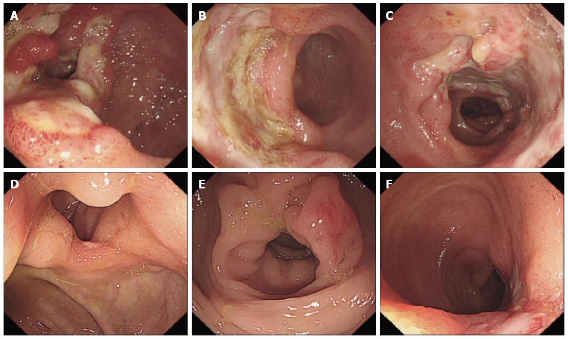
Figure 1 Colonoscopic images of case one.
A-C: Multiple giant and deep ulcers in the ileocecal valve and distal ileum, with polypoid hyperplasia; D-F: Rapid healing of the ulcers in the ileocecal valve and distal ileum and only two healing 2 stage ulcers left.
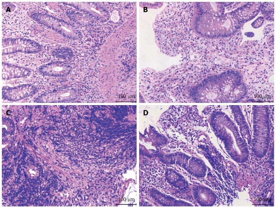
Figure 2 Photomicrograph of biopsy specimens.
A and B: Biopsy specimens of ulcers from case one; C and D: Biopsy specimens of ulcers from case two. Hematoxylin-eosin staining, magnification × 200.
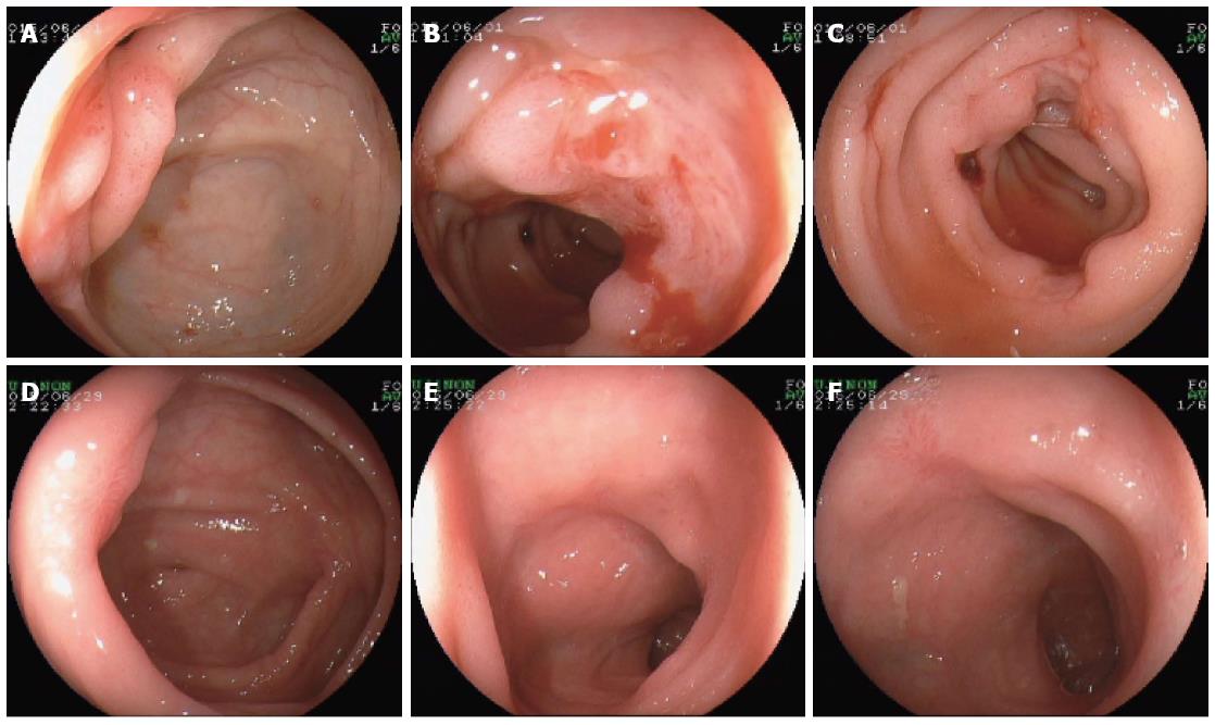
Figure 3 Multiple ulcers in ileocecal valve and distal ileum, with massive fresh blood accumulation (A-C); rapid healing of the ulcers with scar tissue in ileocecal valve and distal ileum (D-F).
- Citation: Guo YW, Gu HY, Abassa KK, Lin XY, Wei XQ. Successful treatment of ileal ulcers caused by immunosuppressants in two organ transplant recipients. World J Gastroenterol 2016; 22(24): 5616-5622
- URL: https://www.wjgnet.com/1007-9327/full/v22/i24/5616.htm
- DOI: https://dx.doi.org/10.3748/wjg.v22.i24.5616


