Copyright
©The Author(s) 2016.
World J Gastroenterol. Jan 14, 2016; 22(2): 874-886
Published online Jan 14, 2016. doi: 10.3748/wjg.v22.i2.874
Published online Jan 14, 2016. doi: 10.3748/wjg.v22.i2.874
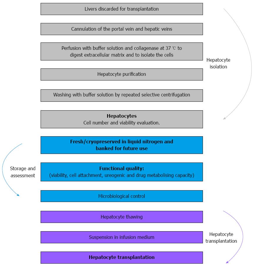
Figure 1 Scheme of human hepatocyte isolation from unused donor liver.
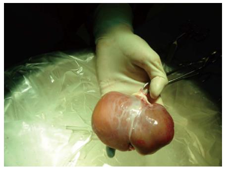
Figure 2 Liver from a neonatal donor to be used for hepatocyte isolation.
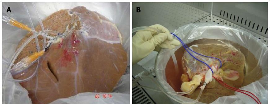
Figure 3 Optimization of the process of hepatocyte isolation.
When livers weighing 1.5-3.5 kg are available, a better recovery was achieved by dividing the hepatic parenchyma (0.5-1 kg) and perfusing each lobe.
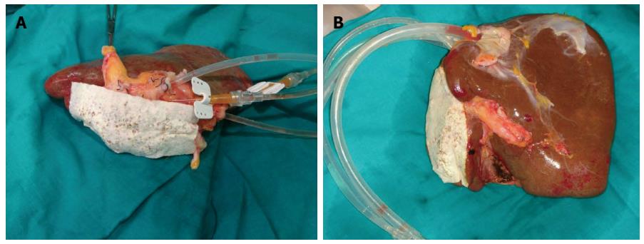
Figure 4 Split liver sutured the arterial, portal and suprahepatic branches in the cut surface.
A: The application TachoSil®, a hemostatic sealant, avoids leakage of the collagenase; B: Cannulation of the hepatic veins allows using bigger calibre cannulas increasing the flow of the perfusates.
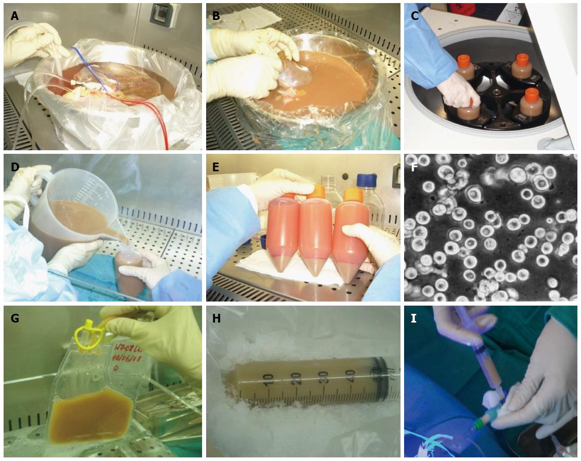
Figure 5 Hepatocyte isolation and transplantation process.
A: Perfusion of the liver at 37 °C; B: Liver after digestion by collagenase perfusion; C: Purification of hepatocytes by low-speed centrifugation; D and E: Hepatocyte suspension is washed twice and pelleted by low-speed centrifugation; F: Freshly isolated hepatocyte suspension; G: Cell thawing by immersing the bags in a fixed-temperature bath at 37 °C; H: Hepatocyte suspension in infusion medium ready to be transplanted; I: Infusion of hepatocytes to the receptor patient.
- Citation: Ibars EP, Cortes M, Tolosa L, Gómez-Lechón MJ, López S, Castell JV, Mir J. Hepatocyte transplantation program: Lessons learned and future strategies. World J Gastroenterol 2016; 22(2): 874-886
- URL: https://www.wjgnet.com/1007-9327/full/v22/i2/874.htm
- DOI: https://dx.doi.org/10.3748/wjg.v22.i2.874


