Copyright
©The Author(s) 2016.
World J Gastroenterol. May 7, 2016; 22(17): 4416-4420
Published online May 7, 2016. doi: 10.3748/wjg.v22.i17.4416
Published online May 7, 2016. doi: 10.3748/wjg.v22.i17.4416
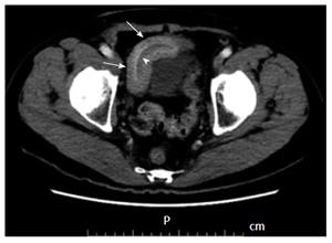
Figure 1 Contrast-enhanced computed tomography images of the lower abdomen.
An abdominal computed tomography showed a thickened small intestinal wall (arrows) and an intraluminal, fat-attenuating lesion in the lower abdomen (arrowhead).
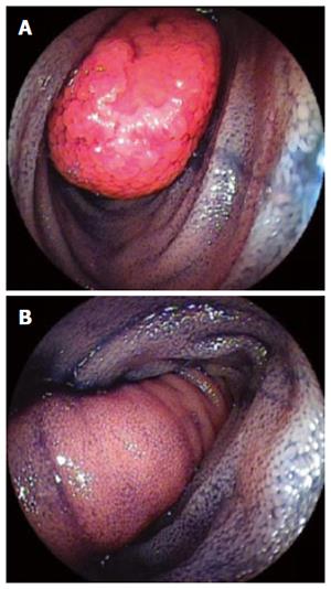
Figure 2 Double-balloon enteroscopy.
A: Double-balloon enteroscopy revealed a sausage-shaped, elongated tumor with a depressed erosion at the tip in the ileum; B: Normal intestinal mucosa was seen on the surface of the tumor.
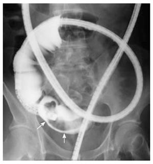
Figure 3 Selective contrast - enhanced radiography.
An elongated, intraluminal polypoid lesion (arrows), approximately 8 cm in size, was seen in the terminal ileum.
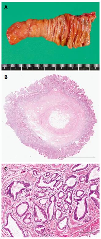
Figure 4 Pathological and histological examination.
A: The opening of the specimen revealed an 8.0 cm × 1.4 cm polypoid lesion; B: Photomicrograph [hematoxylin and eosin (HE) stain] shows that the diverticulum was composed of all layers of the intestinal wall. Scale bars: 6 mm; C: Photomicrograph (HE stain) shows heterotopic gastric mucosa at the tip of the polypoid lesion. Scale bars: 600 μm.
- Citation: Takagaki K, Osawa S, Ito T, Iwaizumi M, Hamaya Y, Tsukui H, Furuta T, Wada H, Baba S, Sugimoto K. Inverted Meckel’s diverticulum preoperatively diagnosed using double-balloon enteroscopy. World J Gastroenterol 2016; 22(17): 4416-4420
- URL: https://www.wjgnet.com/1007-9327/full/v22/i17/4416.htm
- DOI: https://dx.doi.org/10.3748/wjg.v22.i17.4416


