Copyright
©The Author(s) 2016.
World J Gastroenterol. Apr 28, 2016; 22(16): 4270-4274
Published online Apr 28, 2016. doi: 10.3748/wjg.v22.i16.4270
Published online Apr 28, 2016. doi: 10.3748/wjg.v22.i16.4270
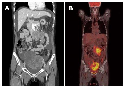
Figure 1 A huge heterogeneous enhancing ovarian mass and peritoneal metastatic lesions were observed.
A: Computed tomography (CT) scan; B: Positron emission tomography-CT scan.
Figure 2 No grossly abnormal lesion was observed in gastroscopy.
A: Examination at first visit to the first hospital; B: At repeat visit to the second hospital; C: At the last visit to our hospital. The black arrows indicate the random biopsy sites. Green matter is remnant gastric content.
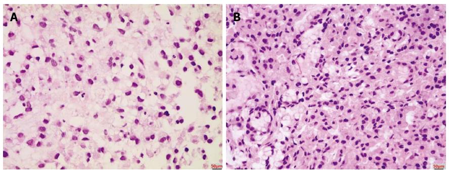
Figure 3 Histopathologic findings.
A: Lymph node needle biopsy; B: Gastric random biopsy; A and B: Hematoxylin-eosin staining and magnification × 400.
- Citation: Lee SH, Lim KH, Song SY, Lee HY, Park SC, Kang CD, Lee SJ, Choi DW, Park SB, Ryu YJ. Occult gastric cancer with distant metastasis proven by random gastric biopsy. World J Gastroenterol 2016; 22(16): 4270-4274
- URL: https://www.wjgnet.com/1007-9327/full/v22/i16/4270.htm
- DOI: https://dx.doi.org/10.3748/wjg.v22.i16.4270


