Copyright
©The Author(s) 2016.
World J Gastroenterol. Apr 21, 2016; 22(15): 3969-3977
Published online Apr 21, 2016. doi: 10.3748/wjg.v22.i15.3969
Published online Apr 21, 2016. doi: 10.3748/wjg.v22.i15.3969
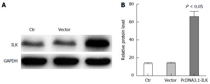
Figure 1 Overexpression of integrin-linked kinase in SW480 cells increased integrin-linked kinase protein expression.
A: Integrin-linked kinase (ILK) expression in the control, vector, and PCDNA3.1-ILK groups were detected by western blot, with GAPDH as an internal control. This experiment was repeated three times; B: Statistical analysis revealed that ILK expressions significantly increased in the PcDNA3.1-ILK group compared with the vector group. GAPDH: Glyceraldehyde-3-phosphate dehydrogenase.
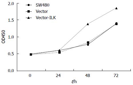
Figure 2 Proliferation rate of SW480 cells due to integrin-linked kinase overexpression.
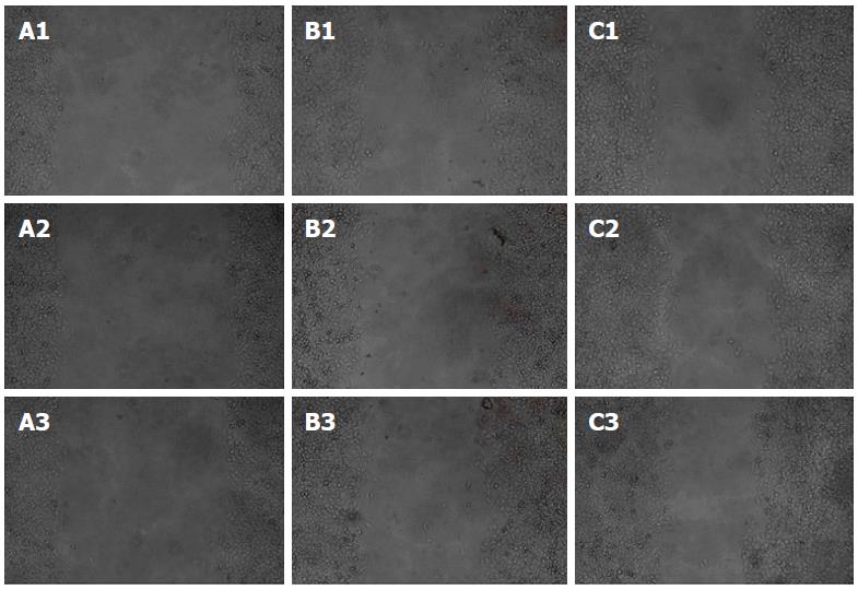
Figure 3 Wound healing assay.
A1, A2, and A3 show the migration condition of cells in the control group after the scrape was made at 0, 6, and 12 h, in which only a small amount of cells migrated after 12 h; B1, B2, and B3 show the migration condition of the vector group, in which only a small amount of cells migrated after 12 h. C1, C2, and C3 show the migration condition of the vector-integrin-linked kinase group, in which a number of cells migrated into the center.
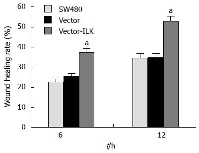
Figure 4 Wound healing rate in the three groups.
aP < 0.05 vs the vector group.
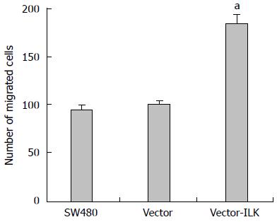
Figure 5 Matrigel invasion assay.
A: The control group, only a small amount of cells migrated to the other side of the chamber; B: The vector group, only a small amount of cells migrated to the other side of the chamber; C: The vector-ILK group, a number of cells migrated to the other side of the chamber.
Figure 6 Number of cells that migrated to the other side of the chamber.
aP < 0.05 vs the vector group.
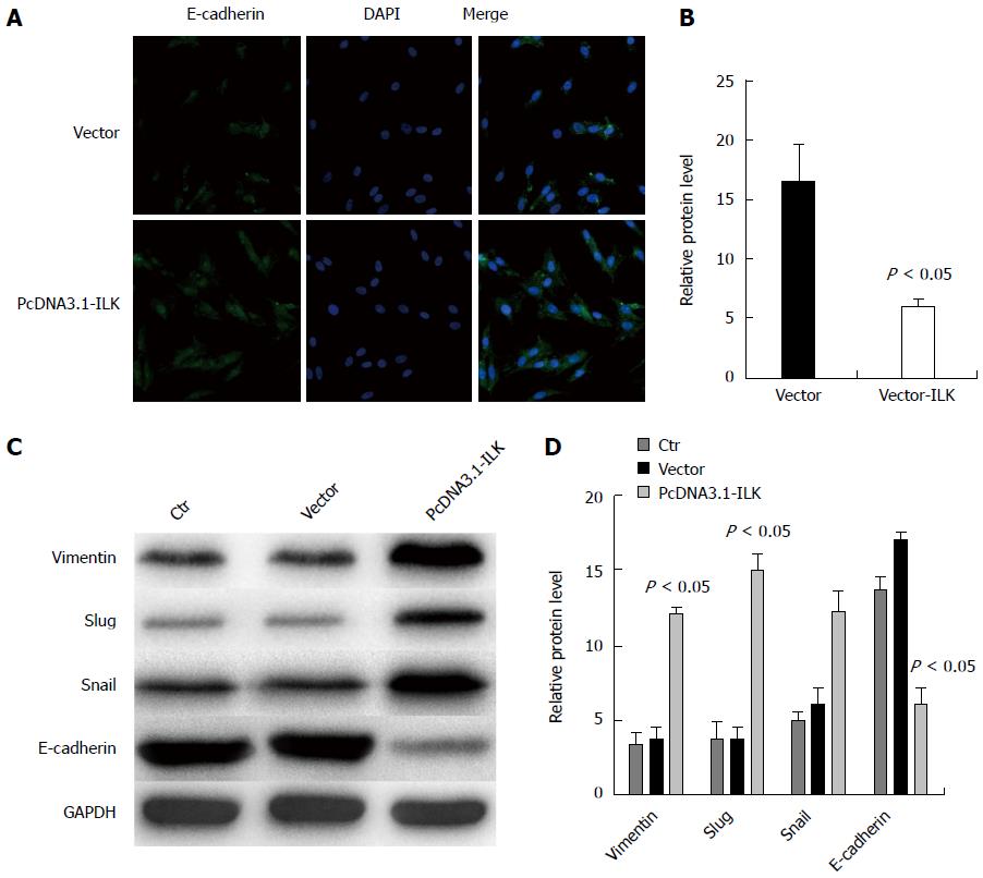
Figure 7 Integrin-linked kinase can promote epithelial-mesenchymal transition occurrence.
A: Immunofluorescence revealed that E-cadherin fluorescence intensity significantly decreased in the pcDNA3.1-ILK group compared with the vector group; B: The difference between the vector group and pcDNA3.1-ILK group was statistically significant; C: Western blot revealed that vimentin, slug, and snail expression was increased, while E-cadherin expressions was reduced in the pcDNA3.1-ILK group; compared with the vector group. This experiment was repeated three times; D: The difference between the vector group and pcDNA3.1-ILK group was statistically significant. ILK: Integrin-linked kinase.
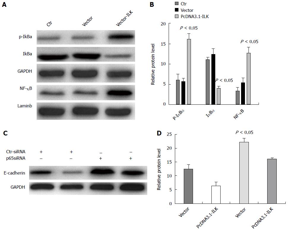
Figure 8 Nuclear factor-κB signaling pathway mediated the integrin-linked kinase-induced epithelial-mesenchymal transition occurrence.
A: Western blot detection of p-IκBa, IγBa, and NF-κB expression; B: Statistical analysis, in which the difference between the vector group and vector-ILK group was statistically significant; C: After treatment with siRNA, E-cadherin expression was detected in cells; D: Statistical analysis: the difference between the vector-ILK cell line in the siRNAp65 treated group and control siRNA group was statistically significant. This experiment was repeated three times. ILK: Integrin-linked kinase; NF-κB: Nuclear factor-κB; siRNA, small interfering RNA.
- Citation: Shen H, Ma JL, Zhang Y, Deng GL, Qu YL, Wu XL, He JX, Zhang S, Zeng S. Integrin-linked kinase overexpression promotes epithelial-mesenchymal transition via nuclear factor-κB signaling in colorectal cancer cells. World J Gastroenterol 2016; 22(15): 3969-3977
- URL: https://www.wjgnet.com/1007-9327/full/v22/i15/3969.htm
- DOI: https://dx.doi.org/10.3748/wjg.v22.i15.3969


