Copyright
©The Author(s) 2015.
World J Gastroenterol. Feb 21, 2015; 21(7): 2254-2259
Published online Feb 21, 2015. doi: 10.3748/wjg.v21.i7.2254
Published online Feb 21, 2015. doi: 10.3748/wjg.v21.i7.2254
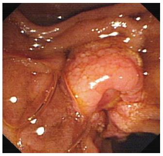
Figure 1 Endoscopic retograde cholangiopancreatography showing a cauliflower-like mass at the duodenal papilla (Case 1).
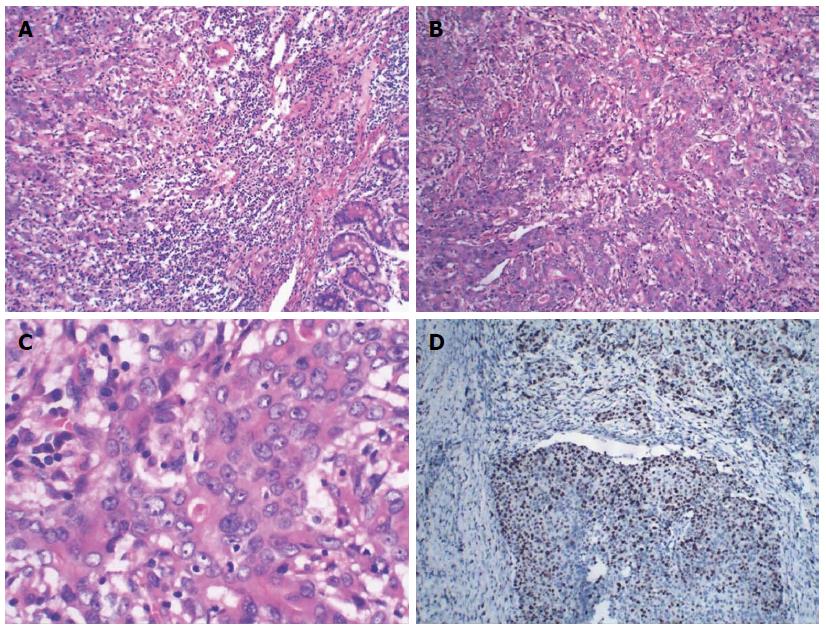
Figure 2 Histological data of Case 1.
A: Poorly differentiated adenocarcinoma admixed with neuroendocrine components [hematoxylin and eosin (HE) staining, × 100]; B: The neuroendocrine component was arranged into a nest and the large cells were consistent in size with round nuclei, abundant cytoplasm and coarse chromatin (HE staining, × 100); C: The neuroendocrine carcinoma cells show high mitotic rate (HE staining, × 400); D: Immunohistochemical assay for Ki67 showing high proliferating index (× 100).
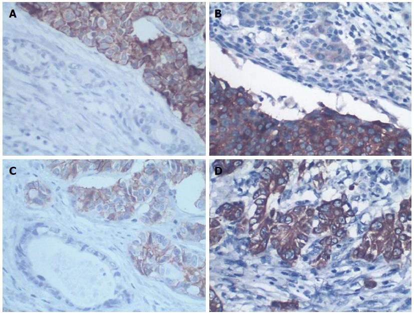
Figure 3 Immunohistochemistry for chromogranin A, synaptophysin, CD56 and CK8 of Case 1.
A: Immunohistochemistry staining for chromogranin A: the neuroendocrine carcinoma components show strong and diffuse cytoplasmic positivity (× 400); B: Immunohistochemistry staining for synaptophysin: the neuroendocrine carcinoma components show strong and diffuse cytoplasmic positivity (× 400); C: Immunohistochemistry staining showed the neuroendocrine carcinoma cells were positive for CD56; D: Immunohistochemistry staining for CK8: moderate and diffuse cytoplasmic positivity of adenocarcinoma cells (× 400).
Figure 4 Histological data of Case 2.
A: Poorly differentiated adenocarcinoma admixed with neuroendocrine components (HE staining, × 100); B: The neuroendocrine components: large cells were characteristic for abundant cytoplasm and nuclear hyperchromatism (HE staining, × 100); C: The neuroendocrine carcinoma cells show high mitotic rate (HE staining, × 400); D: Immunohistochemical assay for Ki67 showing high proliferating index (× 100).
- Citation: Huang Z, Xiao WD, Li Y, Huang S, Cai J, Ao J. Mixed adenoneuroendocrine carcinoma of the ampulla: Two case reports. World J Gastroenterol 2015; 21(7): 2254-2259
- URL: https://www.wjgnet.com/1007-9327/full/v21/i7/2254.htm
- DOI: https://dx.doi.org/10.3748/wjg.v21.i7.2254


