Copyright
©The Author(s) 2015.
World J Gastroenterol. Feb 21, 2015; 21(7): 2225-2228
Published online Feb 21, 2015. doi: 10.3748/wjg.v21.i7.2225
Published online Feb 21, 2015. doi: 10.3748/wjg.v21.i7.2225
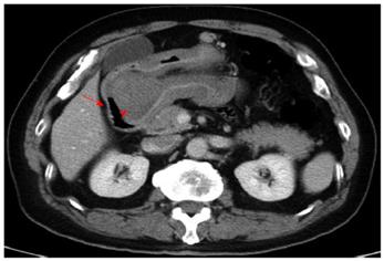
Figure 1 Computed tomography scan revealed a gastroduodenal intussusception of the gastric tumor.
The red arrow marks the duodenum. The red arrowhead marks the tumor in the duodenum.
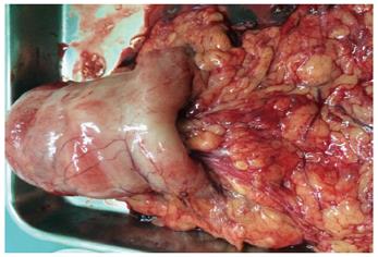
Figure 2 Indentation at the antrum, the origin of intussusception.
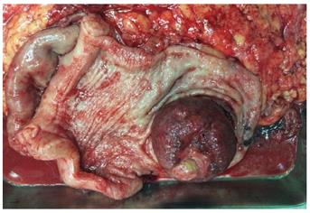
Figure 3 Macroscopic view of the resected specimen shows a lobulated polypoid gastric tumor measuring 50 mm × 50 mm × 48 mm, located at the gastric antrum, which did not infiltrate the mucosa.
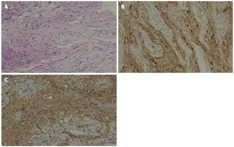
Figure 4 Pathological analysis of the gastric mass confirmed a gastric schwannoma through positive immunohistochemical staining for S-100 protein and vimentin, whereas CD117, CD34, β-catenin, smooth muscle actin, synaptophysin, chromogranin and desmin were negative.
A: The tumor showed typical features of a spindle cell tumor with moderate nuclear pleomorphism but no mitotic activity (original magnification: × 200); B: S-100 protein immunostaining demonstrated strong nuclear and cytoplasmic immunoreactivity; C: Vimentin protein immunostaining demonstrated strong cytoplasmic immunoreactivity.
- Citation: Yang JH, Zhang M, Zhao ZH, Shu Y, Hong J, Cao YJ. Gastroduodenal intussusception due to gastric schwannoma treated by billroth II distal gastrectomy: One case report. World J Gastroenterol 2015; 21(7): 2225-2228
- URL: https://www.wjgnet.com/1007-9327/full/v21/i7/2225.htm
- DOI: https://dx.doi.org/10.3748/wjg.v21.i7.2225


