Copyright
©The Author(s) 2015.
World J Gastroenterol. Feb 21, 2015; 21(7): 2159-2168
Published online Feb 21, 2015. doi: 10.3748/wjg.v21.i7.2159
Published online Feb 21, 2015. doi: 10.3748/wjg.v21.i7.2159
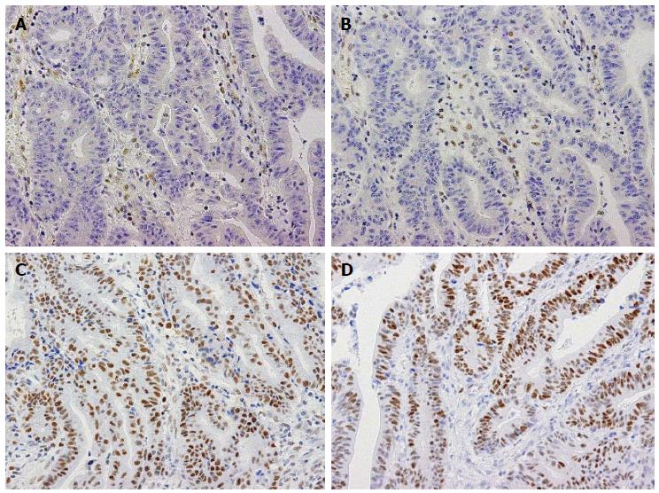
Figure 1 Immunohistochemistry for mismatch repair proteins in a case of gastric cancer with a mismatch repair-deficient phenotype.
Immunohistochemistry for MLH1 (A), PMS2 (B), MSH2 (C), and MSH6 (D). MLH1 and PMS2 expression are absent in tumor cells, whereas stromal cells show nuclear expression (A, B). On the other hand, the tumor cells retain MSH2 and MSH6 expression (C, D). These staining patterns were consistent with those caused by MLH1 deficiency.
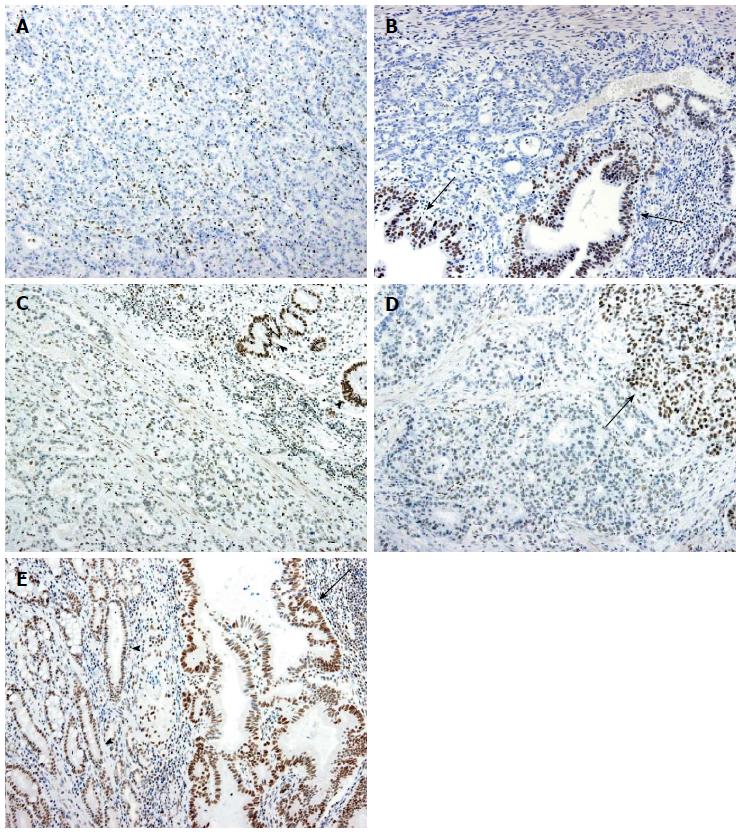
Figure 2 Immunohistochemistry for ARID1A.
A: Homogeneous loss of expression. All the tumor cells show no expression, whereas the stromal cells retain the nuclear expression of ARID1A; B: Heterogeneous loss of expression. Most of the tumor cells show no expression, whereas some of the gland-forming tumor cells retain nuclear expression (arrows); C: Homogeneously weak expression. The tumor cells show the reduced expression of ARID1A. Non-neoplastic gastric glandular cells retain the expression (arrowheads); D: Heterogeneously weak expression. Most of the tumor cells exhibit reduced expression, but a subset of tumor cells retain nuclear expression (arrow); E: Retained expression: Tumor cells (arrow) show strong nuclear ARID1A expression, similar to non-neoplastic glandular cells (arrowheads).
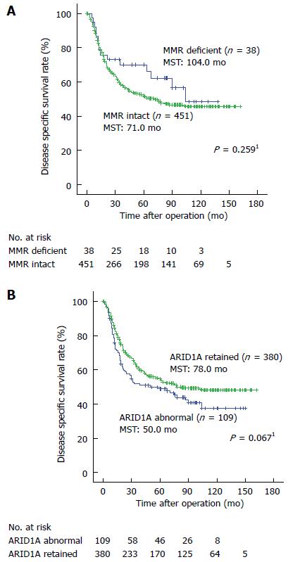
Figure 3 Kaplan-Meier estimates of disease specific survival for patients with gastric cancer according to the mismatch repair (A) and ARID1A expression (B) statuses.
The disease specific survival curves according to the MMR and ARID1A expression statuses did not show significant differences. 1The log-rank test. MST: Median survival time; MMR: Mismatch repair.
- Citation: Inada R, Sekine S, Taniguchi H, Tsuda H, Katai H, Fujiwara T, Kushima R. ARID1A expression in gastric adenocarcinoma: Clinicopathological significance and correlation with DNA mismatch repair status. World J Gastroenterol 2015; 21(7): 2159-2168
- URL: https://www.wjgnet.com/1007-9327/full/v21/i7/2159.htm
- DOI: https://dx.doi.org/10.3748/wjg.v21.i7.2159


