Copyright
©The Author(s) 2015.
World J Gastroenterol. Feb 14, 2015; 21(6): 1857-1864
Published online Feb 14, 2015. doi: 10.3748/wjg.v21.i6.1857
Published online Feb 14, 2015. doi: 10.3748/wjg.v21.i6.1857
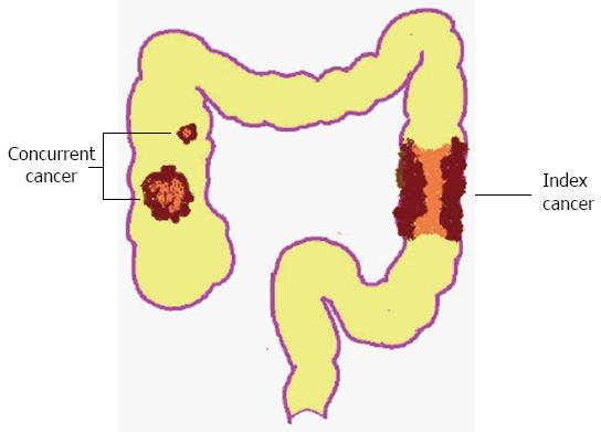
Figure 1 Illustrated model of synchronous colorectal cancers.
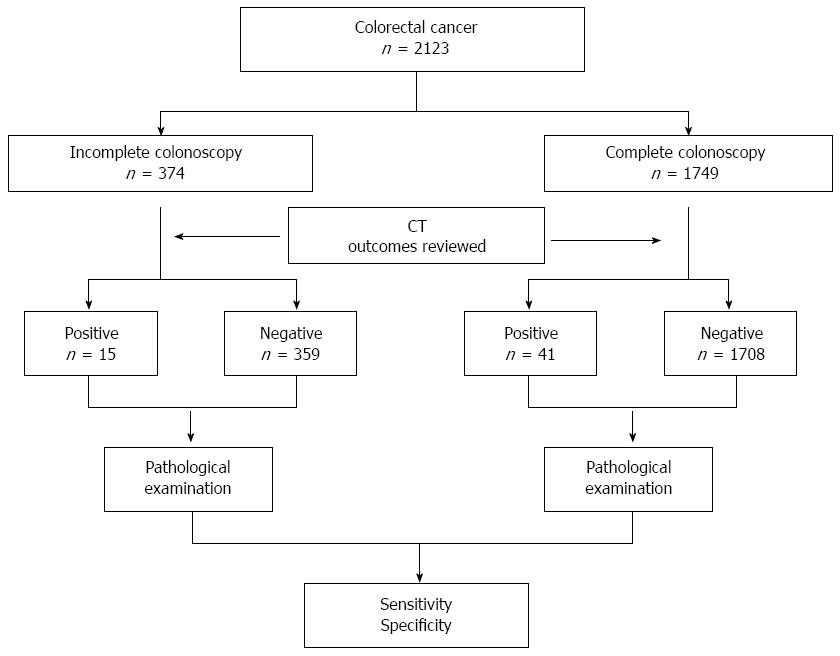
Figure 2 Flow diagram of the present study.
“Positive” refers to synchronous colorectal cancers (SCRCs) detected by computed tomography (CT), while “negative” refers to SCRCs not detected by CT.
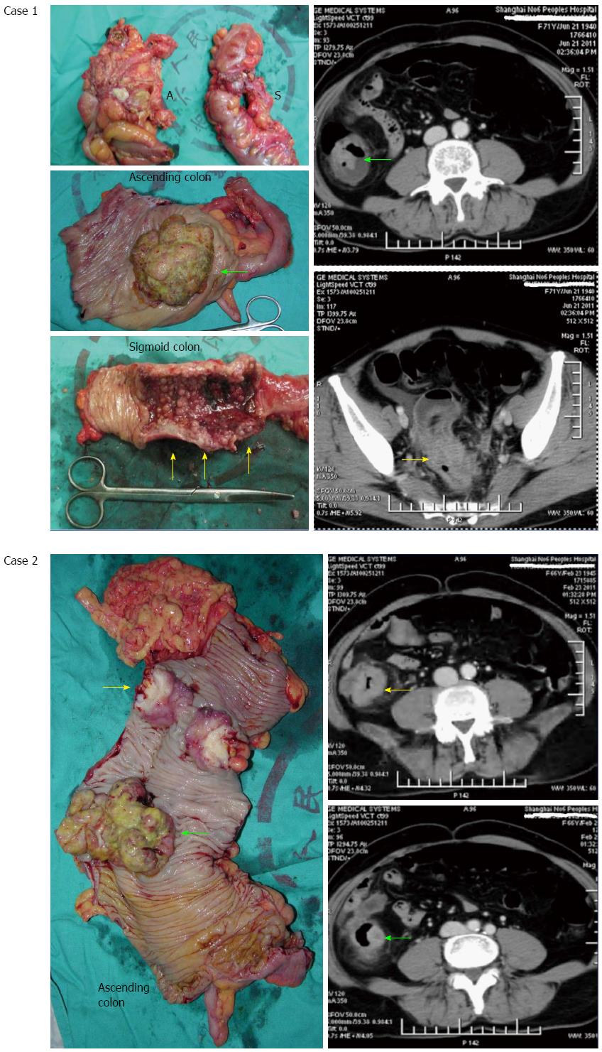
Figure 3 More than 60% of these concurrent cancers were missed by computed tomography.
Case 1: A 56-year-old woman with both index and concurrent cancers located in different bowel segments that were detected by computed tomography (CT). Colonoscopy was hindered by the presence of sigmoid cancer. “A” shows the ascending colon and “S” is the sigmoid colon. The green arrows indicate the concurrent cancer and the yellow arrows indicate the index cancer; Case 2: A 68-year-old man with an index and concurrent cancer both located in the ascending colon. This case was misdiagnosed by CT as a solitary cancer due to the proximity of the two lesions. In addition, colonoscopy was hindered by the presence of a scirrhous carcinoma (index cancer indicated with yellow arrows). The green arrows indicate the concurrent cancer.
- Citation: Pang EJ, Liu WJ, Peng JY, Chen NW, Deng JH. Prediction of synchronous colorectal cancers by computed tomography in subjects receiving an incomplete colonoscopy: A single-center study. World J Gastroenterol 2015; 21(6): 1857-1864
- URL: https://www.wjgnet.com/1007-9327/full/v21/i6/1857.htm
- DOI: https://dx.doi.org/10.3748/wjg.v21.i6.1857


