Copyright
©The Author(s) 2015.
World J Gastroenterol. Feb 14, 2015; 21(6): 1814-1820
Published online Feb 14, 2015. doi: 10.3748/wjg.v21.i6.1814
Published online Feb 14, 2015. doi: 10.3748/wjg.v21.i6.1814
Figure 1 Overexpression of Csk-binding protein decreases the expression of Src in TE-1 cells.
Western blot analysis of the expression of Csk-binding protein (CBP) and Src in TE-1 cells before and after transduction with CBP lentiviral vectors. GAPDH was used as a protein loading control. The expression of CBP was much higher in transduced TE-1 cells than in normal TE-1 and TE-1/pLenO-DCE cells (P < 0.05), and expression of Src was much lower in transduced TE-1 cells than in normal TE-1 and TE-1/pLenO-DCE cells (P < 0.05). 1 = normal TE-1 cells; 2 = TE-1 cells after transfection with pLenO-DCE-(-); 3 = TE-1 cells after overexpression CBP.
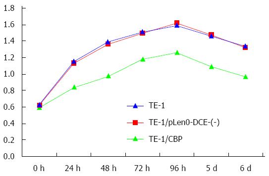
Figure 2 Cell growth of TE-1, TE-1/pLenO-DCE-(-) and TE-1/Csk-binding protein cells.
The growth of TE-1/CBP cells was slower than that of TE-1 and TE-1/pLenO-DCE-(-) cells (P < 0.05).
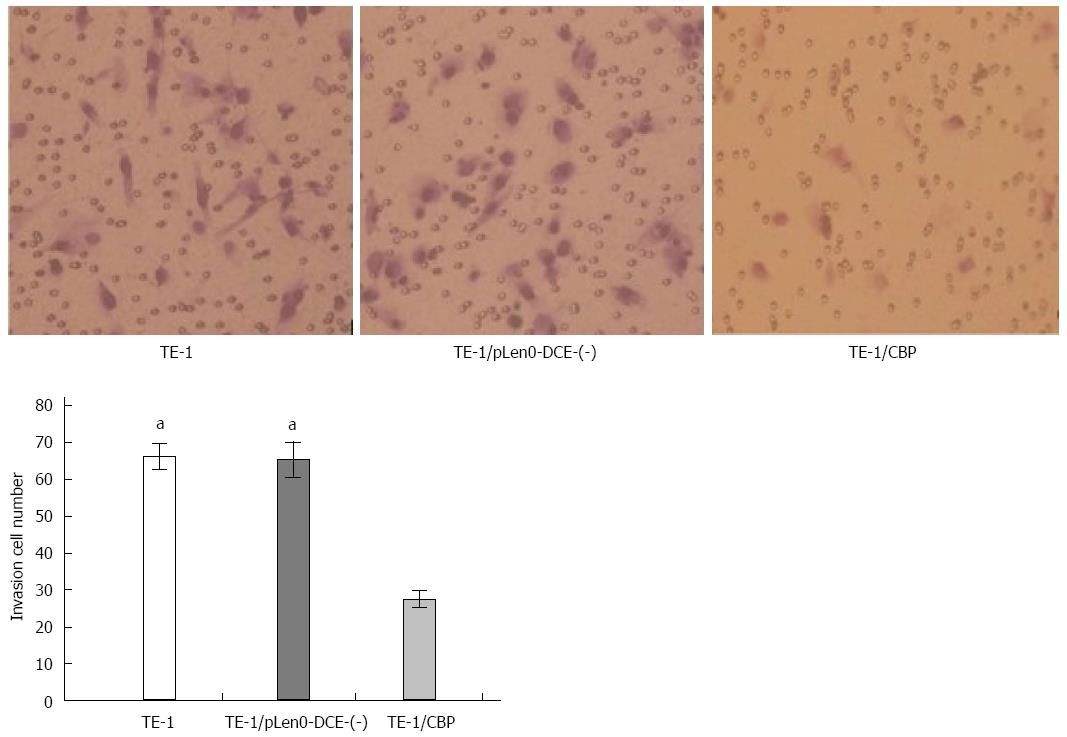
Figure 3 Invasion of TE-1, TE-1/pLenO-DCE-(-) and TE-1/Csk-binding protein cells.
Trypan blue staining showed that TE-1, TE-1/pLenO-DCE-(-) and TE-1/CBP cells passed through the Matrigel (200×). aP < 0.05 vs TE-1/CBP cells, no significant difference compared with TE-1/pLenO-DCE-(-) cells.
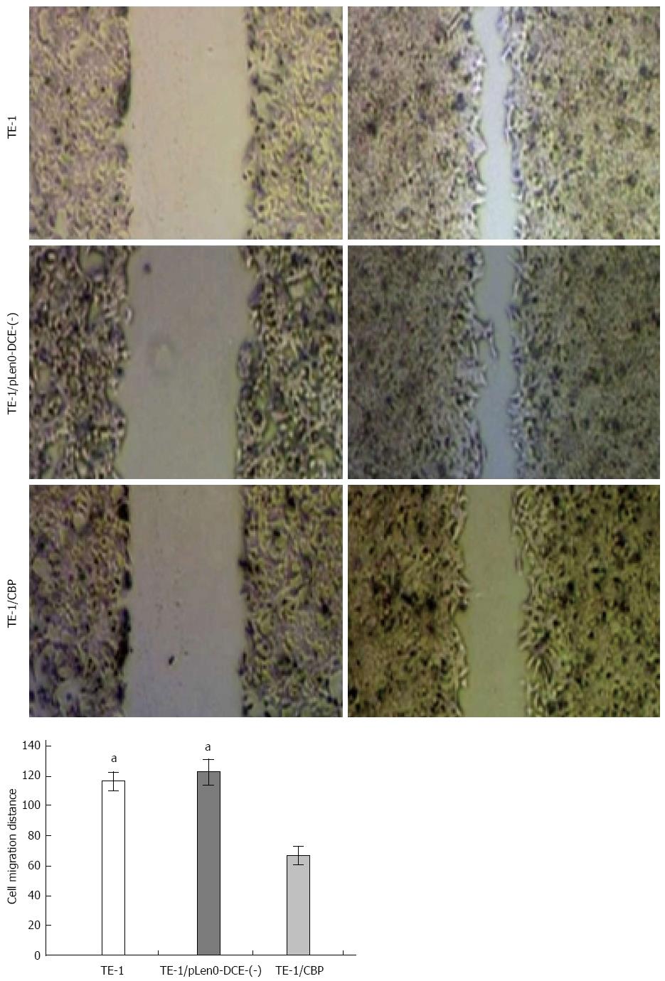
Figure 4 Migration of TE-1, TE-1/pLenO-DCE-(-) and TE-1/ Csk-binding protein cells.
The scratch assay showed that TE-1, TE-1/pLenO-DCE-(-) and TE-1/CBP cells moved into the scratch (200×). The distance of cell migration was assessed. aP < 0.05 vs TE-1/CBP cells, no significant difference compared with TE-1/pLenO-DCE-(-) cells.
- Citation: Zhou D, Dong P, Li YM, Guo FC, Zhang AP, Song RZ, Zhang YM, Li ZY, Yuan D, Yang C. Overexpression of Csk-binding protein decreases growth, invasion, and migration of esophageal carcinoma cells by controlling Src activation. World J Gastroenterol 2015; 21(6): 1814-1820
- URL: https://www.wjgnet.com/1007-9327/full/v21/i6/1814.htm
- DOI: https://dx.doi.org/10.3748/wjg.v21.i6.1814


