Copyright
©The Author(s) 2015.
World J Gastroenterol. Feb 14, 2015; 21(6): 1804-1813
Published online Feb 14, 2015. doi: 10.3748/wjg.v21.i6.1804
Published online Feb 14, 2015. doi: 10.3748/wjg.v21.i6.1804
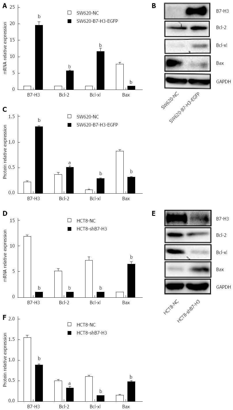
Figure 1 Overexpression of B7-H3 inhibits apoptosis.
A: Real-time PCR for RNA levels of B7-H3, Bcl-2, Bcl-xl and Bax relative to β-actin in stably transfected SW620 cells, control cells (SW620-NC) and B7-H3 overexpressing cells (SW620-B7-H3-EGFP); B: Western blot analysis for B7-H3, Bcl-2, Bcl-xl, Bax and GAPDH protein levels in whole-cell lysates from the SW620 cells; C: Comparison of relative protein levels between the SW620 cells from (B); D: Real-time PCR for RNA levels of B7-H3, Bcl-2, Bcl-xl and Bax relative to β-actin in stably transfected HCT8 cell lines, the control cells (HCT8-NC) and the B7-H3 knockdown cells (HCT8-shB7-H3); E: Western blot analysis for B7-H3, Bcl-2, Bcl-xl, Bax and GAPDH protein levels in whole-cell lysates from the HCT8 cells; F: Comparison of relative protein levels between the HCT8 cells from (E). aP < 0.05, bP < 0.01 vs control. Bax: Bcl-2-associated X protein; Bcl-2: B-cell CLL/lymphoma 2; Bcl-xl: B-cell lymphoma-extra large; NC: Negative control.
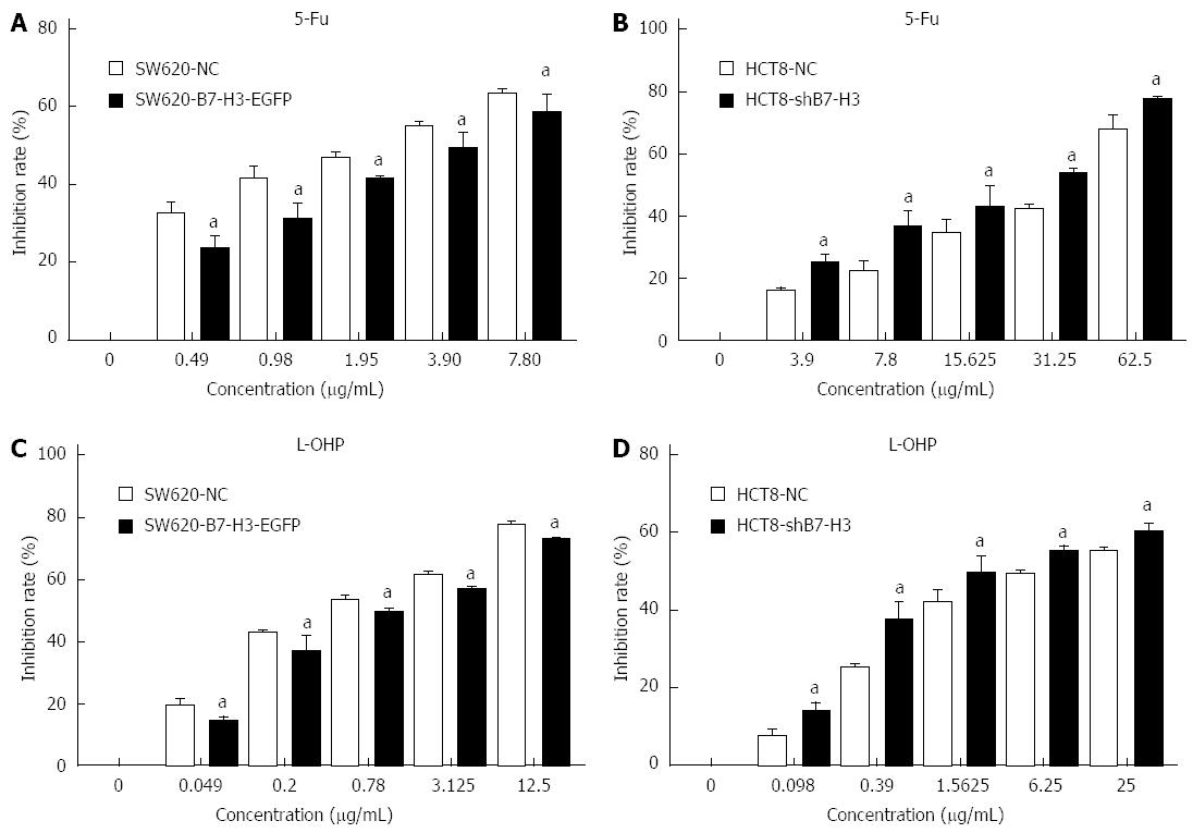
Figure 2 Overexpression of B7-H3 increases cell survival.
Stably transfected SW620 and HCT8 cell lines were incubated with 5-Fu or L-OHP for 48 h. A CCK-8 assay was used to detect the inhibition rate of cells treated with different concentration of different drugs. A: The control cells (SW620-NC) and the B7-H3 overexpressing cells (SW620-B7-H3-EGFP) were incubated with 5-Fu; B: The control cells (HCT8-NC) and the B7-H3 knockdown cells (HCT8-shB7-H3) were incubated with 5-Fu; C: SW620-NC and SW620-B7-H3-EGFP were incubated with L-OHP; D: HCT8-NC and HCT8-shB7-H3 were incubated with L-OHP. aP < 0.05 vs control. 5-Fu: 5-Fluorouracil; L-OHP: Oxaliplatin; NC: Negative control.
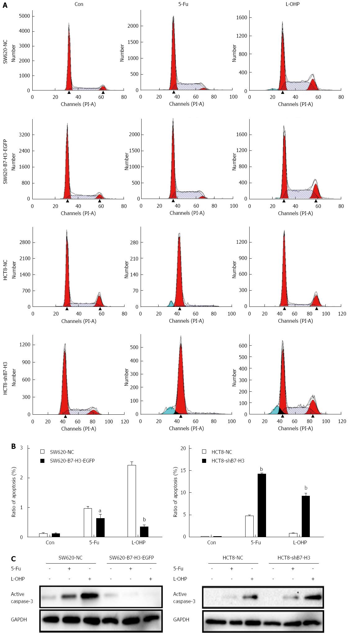
Figure 3 Overexpression of B7-H3 suppresses apoptosis in colorectal cancer cells by weakening the sensitivity to drugs.
Stably transfected SW620 and HCT8 cell lines were incubated with a high concentration of 5-Fu or L-OHP (50 μg/mL) for 48 h. A: The ratio of apoptosis was detected by cell cycle assay according to sub-G1 peak; B: Statistical results were used to analyze the rate of apoptosis from (A); C: Western blot analysis for active Caspase-3 and GAPDH protein levels in whole-cell lysates from stably transfected SW620 and HCT8 cell lines. aP < 0.05, bP < 0.01 vs control. 5-Fu: 5-Fluorouracil; L-OHP: Oxaliplatin.
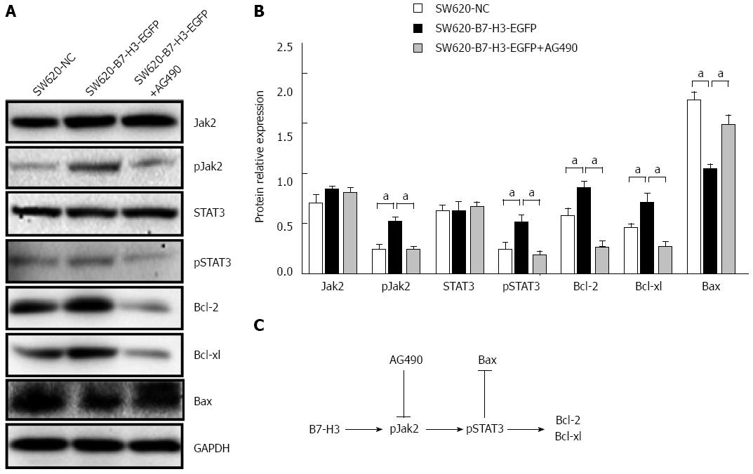
Figure 4 Overexpression of B7-H3 enhances the anti-apoptotic effect in colorectal cancer cells via the activation of the Jak2-STAT3 pathway.
A: Western blot analysis with the control cells (SW620-NC), the B7-H3 overexpressing cells (SW620-B7-H3-EGFP) and the AG490 treated B7-H3 overexpressing cells (SW620-B7-H3-EGFP+AG490) demonstrating the expression of Jak2-STAT3 pathway proteins and apoptosis regulator proteins; B: Comparison of relative protein levels from (A); C: A simple pathway map for B7-H3 regulation of the anti-apoptotic effect in CRC cells. aP < 0.05 vs control. CRC: Colorectal cancer; Jak2: Janus kinase 2; NC: Negative control; STAT3: Signal transducer and activator of transcription 3.
- Citation: Zhang T, Jiang B, Zou ST, Liu F, Hua D. Overexpression of B7-H3 augments anti-apoptosis of colorectal cancer cells by Jak2-STAT3. World J Gastroenterol 2015; 21(6): 1804-1813
- URL: https://www.wjgnet.com/1007-9327/full/v21/i6/1804.htm
- DOI: https://dx.doi.org/10.3748/wjg.v21.i6.1804


