Copyright
©The Author(s) 2015.
World J Gastroenterol. Feb 14, 2015; 21(6): 1759-1764
Published online Feb 14, 2015. doi: 10.3748/wjg.v21.i6.1759
Published online Feb 14, 2015. doi: 10.3748/wjg.v21.i6.1759
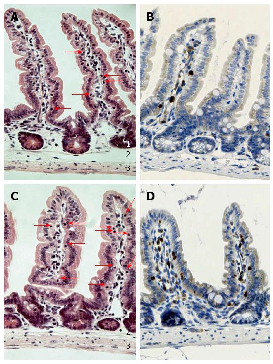
Figure 1 Number of intra-epithelial lymphocytes is diminished in beta-7 integrin-deficient mice.
Histological sections of the jejunum from beta-7 integrin-deficient mice (A and B) as well as age-matched male C57BL/J J mice (C and D) are shown. In the HE stainings (A and C) characteristic intra-epithelial lymphocytes are highlighted by arrows. Anti-CD3 immunostainings are shown (B and D). Original magnification, × 400. 1: Tunica mucosa; 2: Tela submucosa; 3: Muscularis propria.
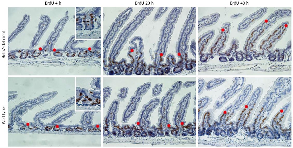
Figure 2 Morphometrical approach for evaluation of enterocyte migration.
Formalin-fixed and paraffin-embedded small intestinal tissues of beta-7 integrin-deficient mice or wild type mice were anti-BrdU immunostained. BrdU was injected 4, 20, or 40 h before sacrificing mice. Red dots illustrate examples of cellmax location. The insets at 4 h illustrate staining results. Original magnification, × 200.
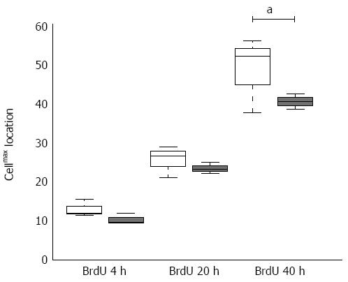
Figure 3 Enterocyte migration is accelerated in beta-7 integrin-deficient mice.
Cellmax locations summarized from duodenum, jejunum, and ileum of beta-7 integrin-deficient mice (white) and wild type littermates (black) 4, 20, 40 h after BrdU application (three animals per group). aP < 0.05 between group.
- Citation: Kaemmerer E, Kuhn P, Schneider U, Clahsen T, Jeon MK, Klaus C, Andruszkow J, Härer M, Ernst S, Schippers A, Wagner N, Gassler N. Beta-7 integrin controls enterocyte migration in the small intestine. World J Gastroenterol 2015; 21(6): 1759-1764
- URL: https://www.wjgnet.com/1007-9327/full/v21/i6/1759.htm
- DOI: https://dx.doi.org/10.3748/wjg.v21.i6.1759


