Copyright
©The Author(s) 2015.
World J Gastroenterol. Dec 14, 2015; 21(46): 13101-13112
Published online Dec 14, 2015. doi: 10.3748/wjg.v21.i46.13101
Published online Dec 14, 2015. doi: 10.3748/wjg.v21.i46.13101
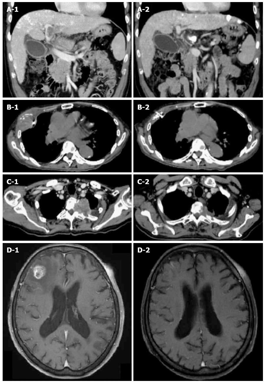
Figure 1 Tumor responses treated with Cyberknife.
A1: CT scan of 59-year-old male with portal vein tumor thrombosis. The tumor is invading the portal vein from the main trunk to the 1st branch. The tumor diameter was 46 mm. A fiducial marker was implanted nearby; A2: Three months after irradiation with 35 Gy/5 fractions. The portal vein tumor thrombosis disappeared completely and the patient achieved CR; B1: CT scan of 85-year-old male with pleural HCC metastasis. The tumor diameter was 53 mm. A fiducial marker was implanted nearby; B2: Three months after irradiation with 30 Gy/5 fractions. The tumor disappeared completely and the patient achieved CR; C1: CT scan of 72-year-old male with thoracic spine HCC metastasis. Tumor is invading the left side of the thoracic spine at T2-3 causing bone destruction. The tumor diameter was 52 mm; C2: Three months after irradiation with 30 Gy/5 fractions. The tumor decreased to 33 mm (37% reduction in size) and the patient achieved PR; D1: T1-weighted MRI of 83-year-old female with brain HCC metastasis. There is a right frontal lobe lesion with gadolinium enhancement. The tumor diameter was 19 mm; D2: One month after irradiation of 20 Gy/1 fraction. The tumor disappeared completely and the patient achieved CR. CR: Complete response; CT: Computed tomography; HCC: Hepatocellular carcinoma.
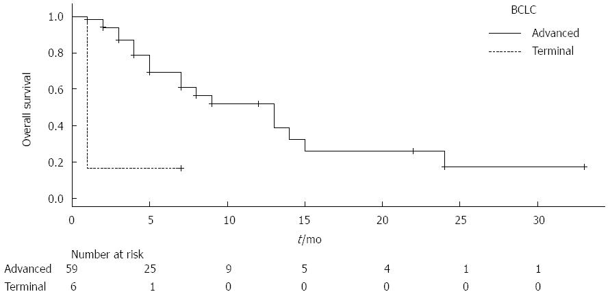
Figure 2 Kaplan Meier curves for overall survival.
The median survival times for the advanced stage patients and terminal stage patients were 13 mo and 1 mo respectively.
- Citation: Kato H, Yoshida H, Taniguch H, Nomura R, Sato K, Suzuki I, Nakata R. Cyberknife treatment for advanced or terminal stage hepatocellular carcinoma. World J Gastroenterol 2015; 21(46): 13101-13112
- URL: https://www.wjgnet.com/1007-9327/full/v21/i46/13101.htm
- DOI: https://dx.doi.org/10.3748/wjg.v21.i46.13101


