Copyright
©The Author(s) 2015.
World J Gastroenterol. Dec 7, 2015; 21(45): 12976-12980
Published online Dec 7, 2015. doi: 10.3748/wjg.v21.i45.12976
Published online Dec 7, 2015. doi: 10.3748/wjg.v21.i45.12976
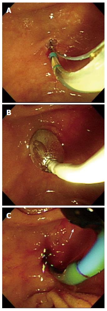
Figure 1 The patient underwent endoscopic retrograde cholangiopancreatography with sphincterotomy and endoscopic papillary balloon dilatation.
A: Endoscopic retrograde cholangiopancreatography with sphincterotomy; B: Endoscopic papillary balloon dilatation (0.8 cm, 8 atm × 3 min); C: Black stone (white arrow) was extracted with a balloon catheter.
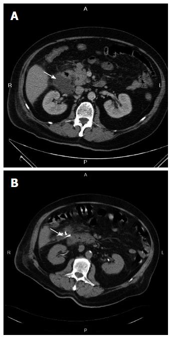
Figure 2 Computed tomography scan.
A: Showing a hypodense abscess (arrow) between the liver and right kidney; B: Showing pig-tail drainage (arrow) of the abscess.
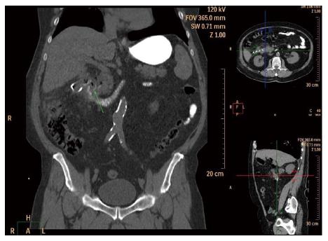
Figure 3 Computed tomography scan of the abdomen.
Green arrow showing perforation site.
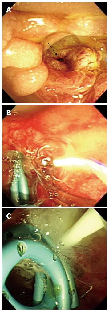
Figure 4 Endoscopic retrograde cholangiopancreatography.
Endoscopic retrograde cholangiopancreatography revealed a perforation (arrow) in the duodenum (A); Two plastic stents were inserted into the endoscopic papillary balloon dilatation before fibrin sealant injection (B); Whitish tissue sealant was injected into the perforation in the right upper corner (C).
- Citation: Yang HY, Chen JH. Endoscopic fibrin sealant closure of duodenal perforation after endoscopic retrograde cholangiopancreatography. World J Gastroenterol 2015; 21(45): 12976-12980
- URL: https://www.wjgnet.com/1007-9327/full/v21/i45/12976.htm
- DOI: https://dx.doi.org/10.3748/wjg.v21.i45.12976


