Copyright
©The Author(s) 2015.
World J Gastroenterol. Dec 7, 2015; 21(45): 12857-12864
Published online Dec 7, 2015. doi: 10.3748/wjg.v21.i45.12857
Published online Dec 7, 2015. doi: 10.3748/wjg.v21.i45.12857
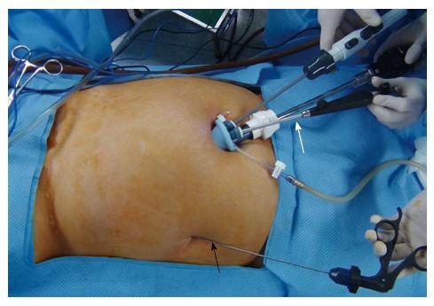
Figure 1 External view of needlescopic grasper-assisted single-incision laparoscopic common bile duct exploration.
The needlescopic grasper (black arrow) was used for traction through a direct puncture on the right abdomen along the right anterior axillary line. The snake liver retractor (white arrow) was used for cephalad traction of the liver to obtain better visualization.
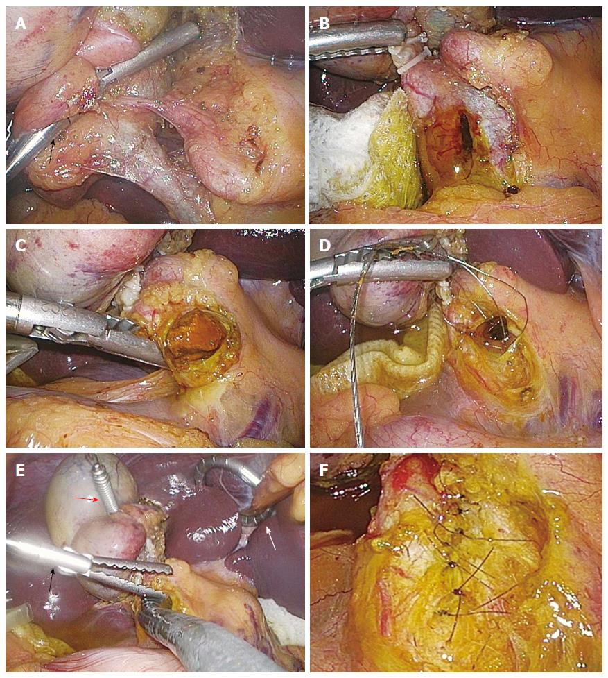
Figure 2 Operative illustrations of needlescopic grasper-assisted single-incision laparoscopic common bile duct exploration.
The needlescopic grasper helped with obtaining a critical view of safety by retracting the gallbladder laterally (A). After ligation and transection of the cystic artery and duct, the common bile duct (CBD) was opened longitudinally using an endoknife (B) The CBD stones were extracted through various methods including direct extraction (C) or the use of a stone basket (D). After extraction of the CBD stones, completion choledochoscopy was performed to check for the presence of remnant stone(s) (E). After CBD clearance, the choledochotomy was primarily closed using either interrupted (F) or running sutures. Black arrows indicates lateral retraction of the gallbladder by the needlescopic grasper. A white and a red arrows indicate the snake retractor and Endograb, respectively.
- Citation: Kim SJ, Kim KH, An CH, Kim JS. Innovative technique of needlescopic grasper-assisted single-incision laparoscopic common bile duct exploration: A comparative study. World J Gastroenterol 2015; 21(45): 12857-12864
- URL: https://www.wjgnet.com/1007-9327/full/v21/i45/12857.htm
- DOI: https://dx.doi.org/10.3748/wjg.v21.i45.12857


