Copyright
©The Author(s) 2015.
World J Gastroenterol. Nov 28, 2015; 21(44): 12729-12734
Published online Nov 28, 2015. doi: 10.3748/wjg.v21.i44.12729
Published online Nov 28, 2015. doi: 10.3748/wjg.v21.i44.12729
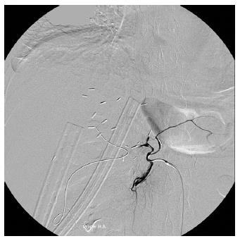
Figure 1 Angiography in a 60-year-old woman 9 d after living donor liver transplantation shows thrombosis at the proper hepatic artery anastomosis, in which endovascular intervention failed to restore blood flow through the hepatic artery occlusion.
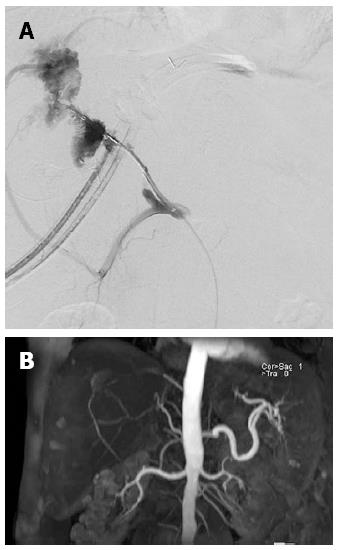
Figure 2 Angiography in a 49-year-old man 8 d after living donor liver transplantation shows persistent thrombosis at the anastomosis site of hepatic artery (A), however magnetic resonance angiography at the 8th month after liver transplantation shows recanalization of intrahepatic artery via right inferior phrenic artery (B).
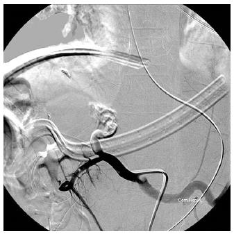
Figure 3 Angiography in a 13-year-old boy 2 dafter living donor liver transplantation shows total occlusion of the proper hepatic artery despite injection of 10000 IU of urokinase through a microcatheter with tip inserted into the thrombosed proper hepatic artery.
- Citation: Hsiao CY, Ho CM, Wu YM, Ho MC, Hu RH, Lee PH. Management of early hepatic artery occlusion after liver transplantation with failed rescue. World J Gastroenterol 2015; 21(44): 12729-12734
- URL: https://www.wjgnet.com/1007-9327/full/v21/i44/12729.htm
- DOI: https://dx.doi.org/10.3748/wjg.v21.i44.12729


