Copyright
©The Author(s) 2015.
World J Gastroenterol. Nov 28, 2015; 21(44): 12722-12728
Published online Nov 28, 2015. doi: 10.3748/wjg.v21.i44.12722
Published online Nov 28, 2015. doi: 10.3748/wjg.v21.i44.12722
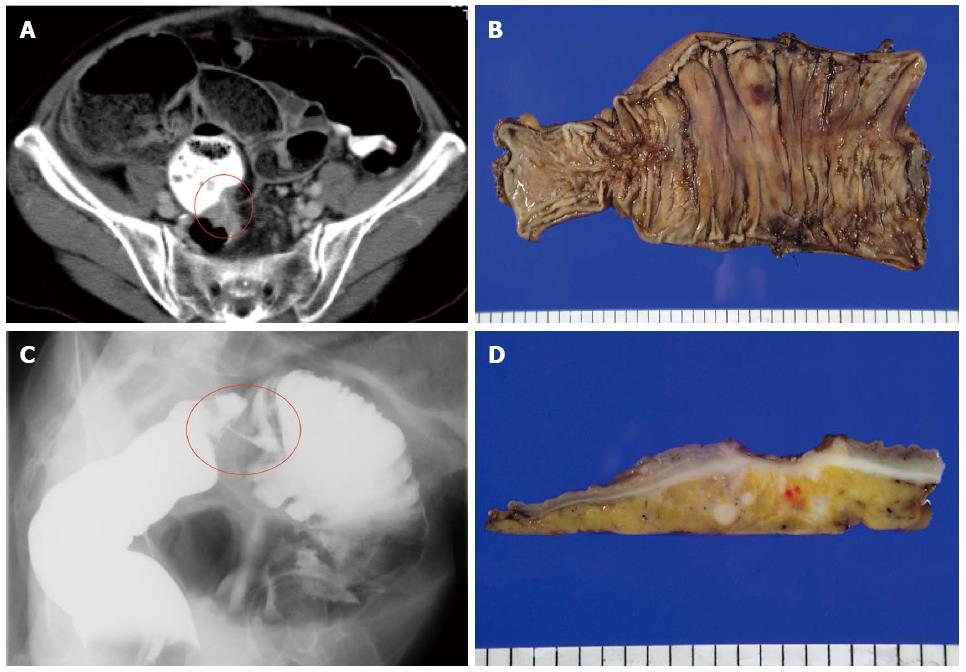
Figure 1 Computed tomography showed hypertrophy and obstruction of rectum and severe colonic dilatation (A); Radiographical examination revealed rectosigmoid obstruction (B); and Histological examination showed moderately differentiated adenocarcinoma, pSS, pN3, pH0, pP1, pM1 (para-aortic lymph node, dissemination) fStage IV (C and D).
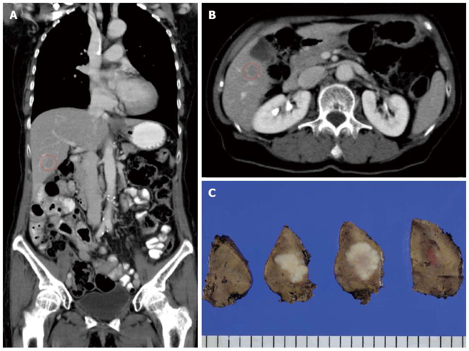
Figure 2 Computed tomography detected an 11 mm of liver metastasis in the posteroinferior segment of right hematic lobe (A and B); and Histological examination revealed moderately differentiated adenocarcinoma as a metastatic rectal cancer with cut end microscopically positive (C).
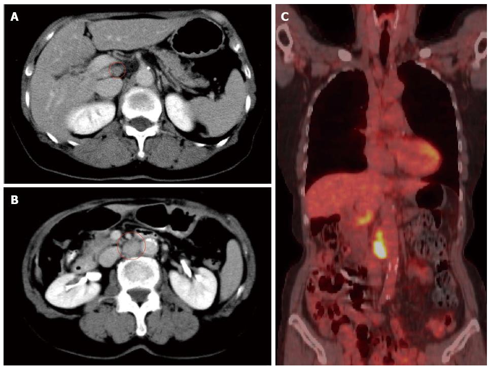
Figure 3 Computed tomography detected a 20 mm of para-aortic lymph node metastasis and a 10 mm of lymph node metastasis at the hepato-duodenal ligament (A and B); and Positron emission tomography was performed and detected the recurrences at the same lesions (C).
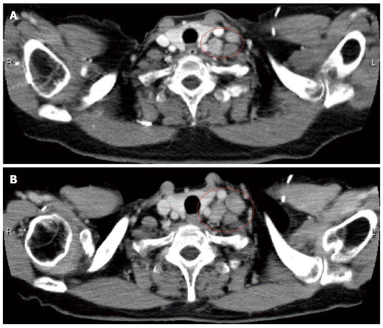
Figure 4 Computed tomography detected two swelling 12 mm of lymph nodes at the left supraclavicular (A); and Virchow lymph nodes had slowly grown up to 17 mm (B).
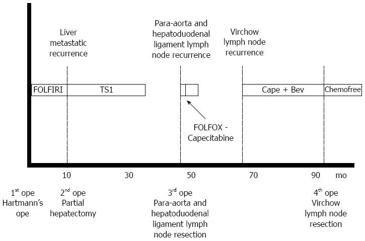
Figure 5 Clinical course of this case.
- Citation: Takeshita N, Fukunaga T, Kimura M, Sugamoto Y, Tasaki K, Hoshino I, Ota T, Maruyama T, Tamachi T, Hosokawa T, Asai Y, Matsubara H. Successful resection of metachronous para-aortic, Virchow lymph node and liver metastatic recurrence of rectal cancer. World J Gastroenterol 2015; 21(44): 12722-12728
- URL: https://www.wjgnet.com/1007-9327/full/v21/i44/12722.htm
- DOI: https://dx.doi.org/10.3748/wjg.v21.i44.12722


