Copyright
©The Author(s) 2015.
World J Gastroenterol. Nov 28, 2015; 21(44): 12593-12604
Published online Nov 28, 2015. doi: 10.3748/wjg.v21.i44.12593
Published online Nov 28, 2015. doi: 10.3748/wjg.v21.i44.12593
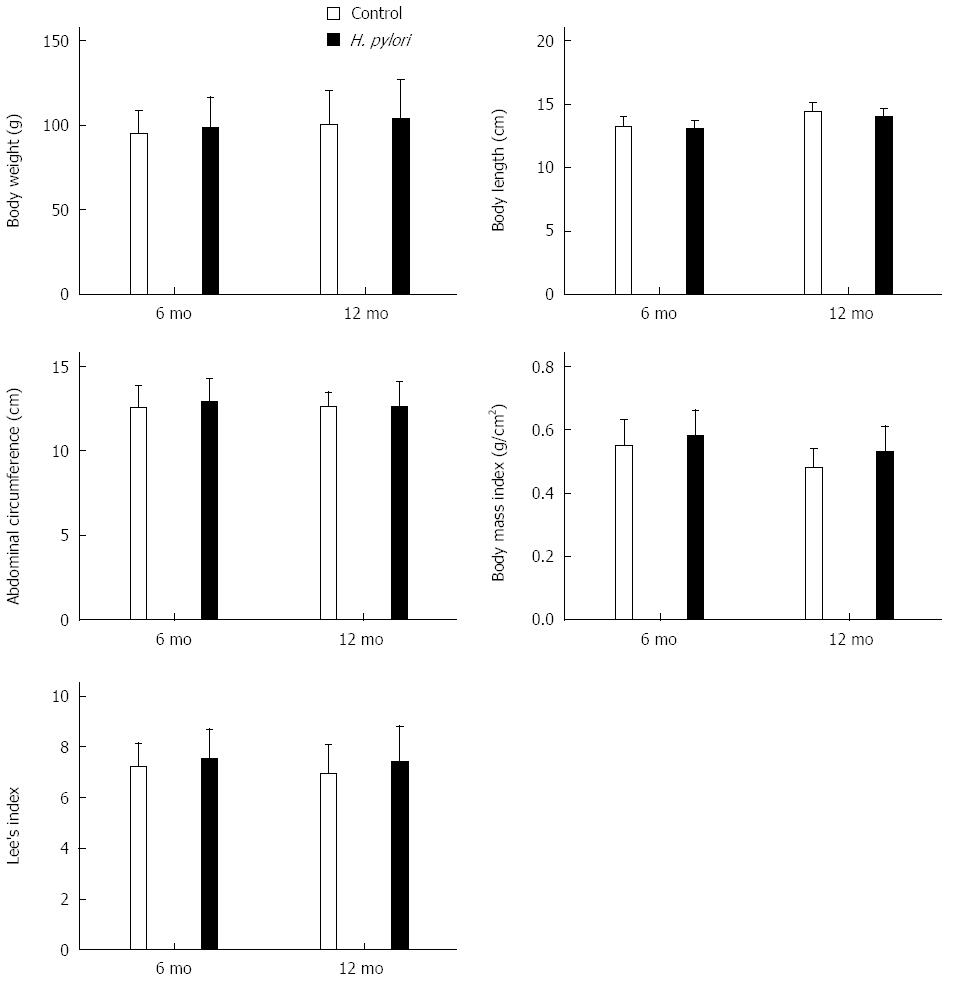
Figure 1 Effects of Helicobacter pylori infection on body indexes of Mongolian gerbils at different time points.
The body weight, body length, and abdominal circumference were measured, and body mass index and Lee’s index were calculated at 6 and 12 mo after Helicobacter pylori (H. pylori) infection. The data denote an upward trend of these parameters, although there were no significant differences (P > 0.05). Bars represent the mean ± SEM, n = 8-10 mice per group.
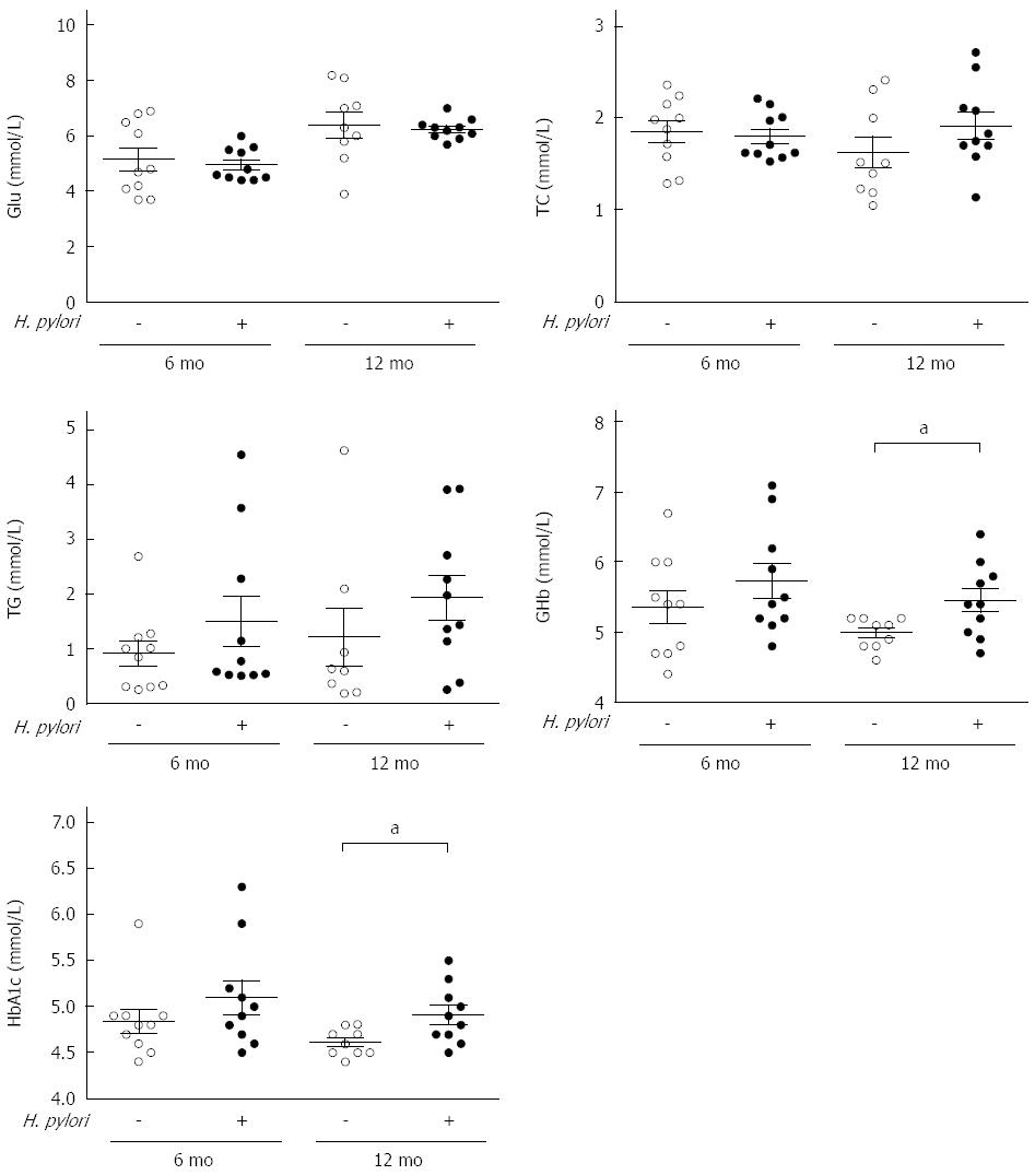
Figure 2 Measurements of serum biochemical parameters of Mongolian gerbils after Helicobacter pylori infection at different time points.
Serum concentration of fasting glucose (Glu), triacylglycerol (TG), total cholesterol (TC), and glycated hemoglobin (GHb) and hemoglobin A1c (HbA1c) were measured at 6 and 12 mo after Helicobacter pylori (H. pylori) infection. Data are presented as mean ± SEM from eight to ten mice per group. aP < 0.05 between H. pylori infected mice and their controls at 12 mo concerning GHb and HbA1c.
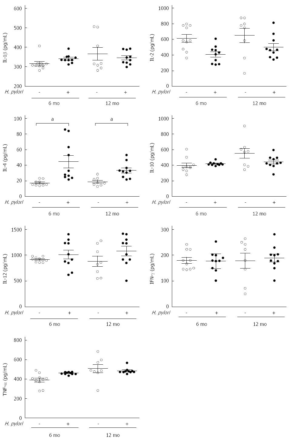
Figure 3 Effects of chronic Helicobacter pylori infection on the serum inflammatory cytokines in Mongolian gerbils at different time points.
The serum levels of cytokines including IL-1β, IL-2, IL-12, IL-4, IL-10, IFN-γ, and TNF-α were measured by ELISA at 6 and 12 mo after Helicobacter pylori (H. pylori) infection. Data are presented as mean ± SEM from eight to ten mice per group. Cytokines were not significant different except IL-4 (aP < 0.05 between H. pylori infected mice and their controls). IL: Interleukin; TNF: Tumor necrosis factor; IFN: Interferon.
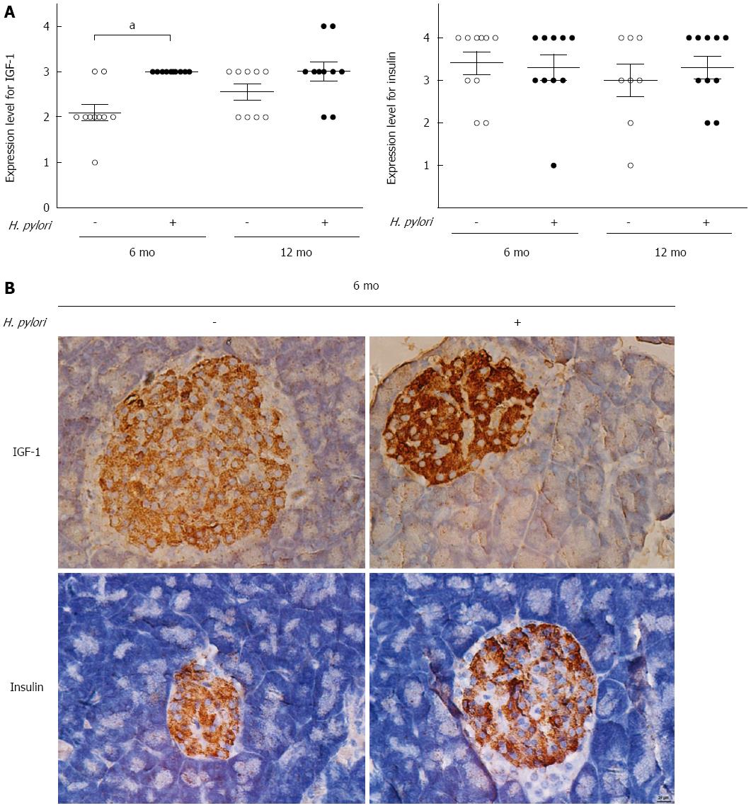
Figure 4 Effects of chronic Helicobacter pylori infection on the expression of insulin-like growth factor-1 and insulin in the pancreas of Mongolian gerbils.
A: Immunohistochemistry scores of insulin-like growth factor-1 (IGF-1) and insulin were determined in the control and Helicobacter pylori (H. pylori) groups at 6 and 12 mo (n = 8-10). Data are expressed as the mean ± SEM. aP < 0.05 between H. pylori infected mice at 6 mo and their controls concerning the expression of IGF-1; B: Representative immunohistochemical staining of IGF-1 and insulin expression in the pancreas of Mongolian gerbils at 6 mo after H. pylori infection (magnification × 400).
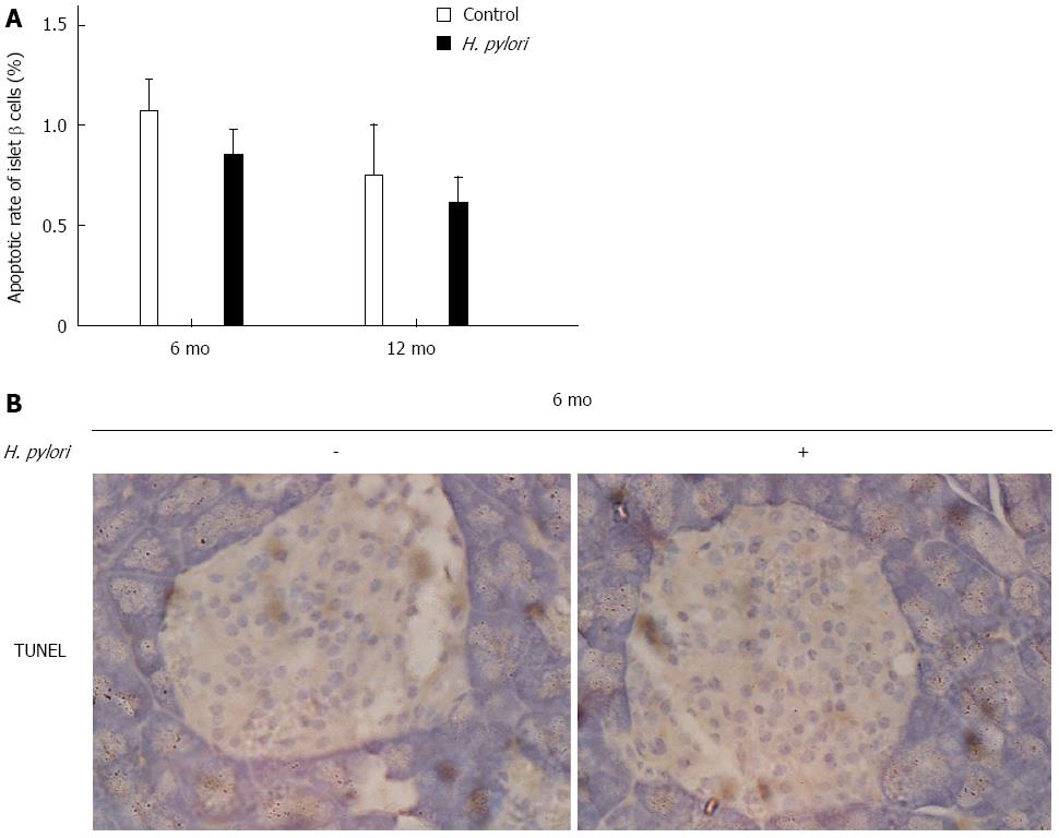
Figure 5 Effects of chronic Helicobacter pylori infection on the apoptosis of islet β-cells in Mongolian gerbils.
A: Apoptotic rate of islet β-cells was detected by TUNEL assay in the control and Helicobacter pylori (H. pylori) groups at 6 and 12 mo. Data are expressed as the means ± SEM (n = 8-10 mice/group). P > 0.05 was observed between H. pylori infected mice and their controls; B: Representative images of islet β-cells in the pancreas of Mongolian gerbils at 6 mo after H. pylori infection by TUNEL staining (magnification × 400).
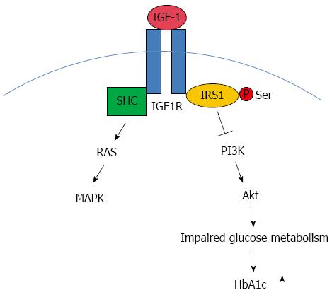
Figure 6 Insulin-like growth factor-1 signaling with regard to Helicobacter pylori associated glucose dysregulation.
Insulin-like growth factor-1 (IGF-1), upon binding to its receptor IGF-1R, activates the intrinsic tyrosine kinase activity. IGF-1R then phosphorylates substrate proteins, including members of the IRS family such as IRS1 and Shc on selective tyrosine residues. Downstream to the receptors are two major pathways: the phosphatidylinositol-3 kinase (PI3K)/Akt pathway and MAPK pathway. IGF-1 acts via the MAPK pathway to mediate growth responses. The PI3K pathway is thought to have predominantly metabolic effects. We supposed that this pathway might play a part in Helicobacter pylori (H. pylori) associated abnormal glucose metabolism and the upregulation of HbA1c. SHC: Spontaneous human combustion.
-
Citation: Yang Z, Li W, He C, Xie C, Zhu Y, Lu NH. Potential effect of chronic
Helicobacter pylori infection on glucose metabolism of Mongolian gerbils. World J Gastroenterol 2015; 21(44): 12593-12604 - URL: https://www.wjgnet.com/1007-9327/full/v21/i44/12593.htm
- DOI: https://dx.doi.org/10.3748/wjg.v21.i44.12593


