Copyright
©The Author(s) 2015.
World J Gastroenterol. Nov 28, 2015; 21(44): 12544-12557
Published online Nov 28, 2015. doi: 10.3748/wjg.v21.i44.12544
Published online Nov 28, 2015. doi: 10.3748/wjg.v21.i44.12544
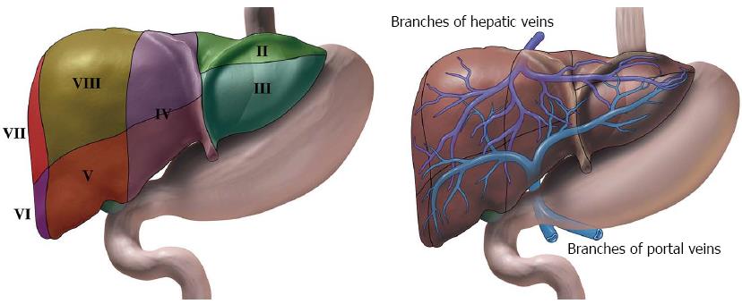
Figure 1 Illustrations of liver and its surrounding stomach and duodenum.
A: The liver can subsequently be divided into 8 segments that is served independently by a secondary or tertiary branch of the portal triad. B: The left hepatic vein divides the left lobe into lateral (II2, III3) and medial (IV4a, IV4b) segments. The right hepatic vein divides the right lobe into anterior (V5, VIII8) and posterior (VI6, VII7) segments. The portal vein divides the liver into upper (II2, IV4a, VIII8, VII7) and lower (III3, IV4b, V5, VI6) segments. The segments are labeled in a clockwise manner. In a normal frontal view segments I1, VI6 and VII7 are not visible.
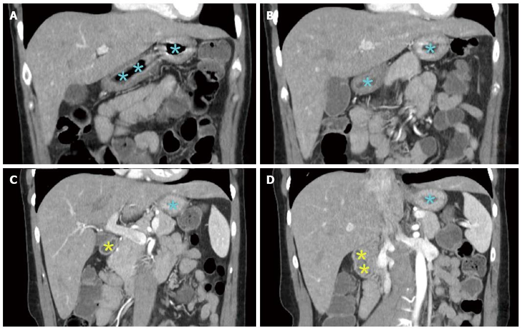
Figure 2 Selected computerized tomography scan images showing the liver and its surrounding stomach and duodenum.
Both the left lobe and caudate lobe lie in close proximity to stomach (blue colored asterisk indicated gastric lumen) and duodenum (yellow colored asterisk indicated bulb and duodenal lumen), hence providing an easy access during endoscopic ultrasonography (EUS). The caudate lobe and gastrohepatic space can be accessed by EUS while are anatomically difficult to approached by trans-abdominal ultrasound. EUS is limited in its ability to access the portion of the right lobe adjacent to the dome of the diaphragm along with its lateral and inferior portions.
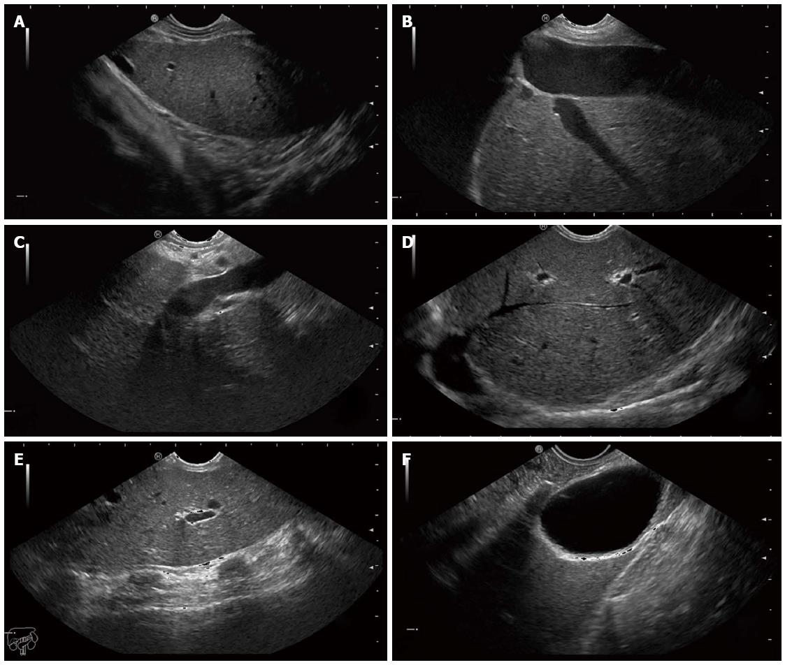
Figure 3 Endoscopic ultrasound images of the hepatic structures with the tip of the linear echoendoscope at different positions.
A: Endoscopic ultrasound (EUS) image of the left liver lobe with the diaphragm. The image is obtained from the cardia region; B: EUS image of the left liver lobe with the inferior vena cava and a hepatic vein; C: EUS image of the liver at the portal ligament region showing from the transducer, the hepatic artery, the portal vein and a short segment of the common bile duct. The transducer is located in the stomach; D: EUS image of the liver looking over the hepatic dome; E: EUS image of the right hepatic lobe. Note the shadows from the ribs at the anterior abdominal wall; F: EUS image of the liver with the gall bladder. The transducer is located in the first part of the duodenum.
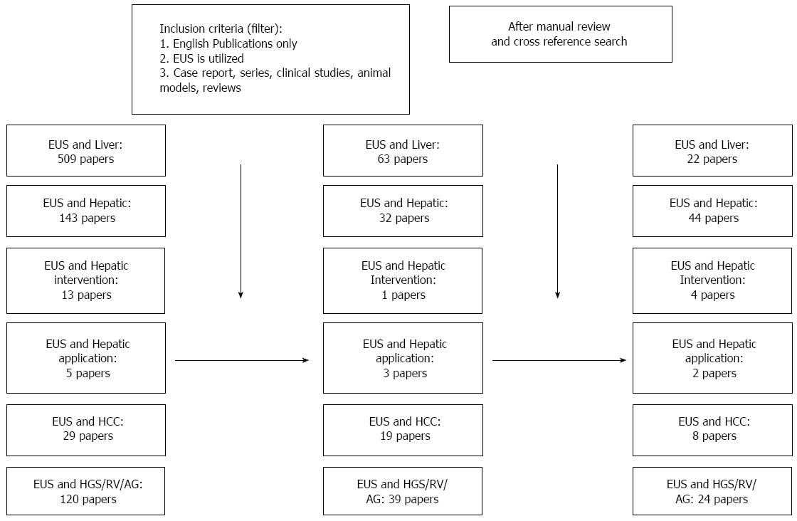
Figure 4 PubMed result search flow sheet.
PubMed search was performed on December 20, 2014. EUS: Endoscopic ultrasound; HCC: Hepatocellular carcinoma; HGS: Hepaticogastrostomy.
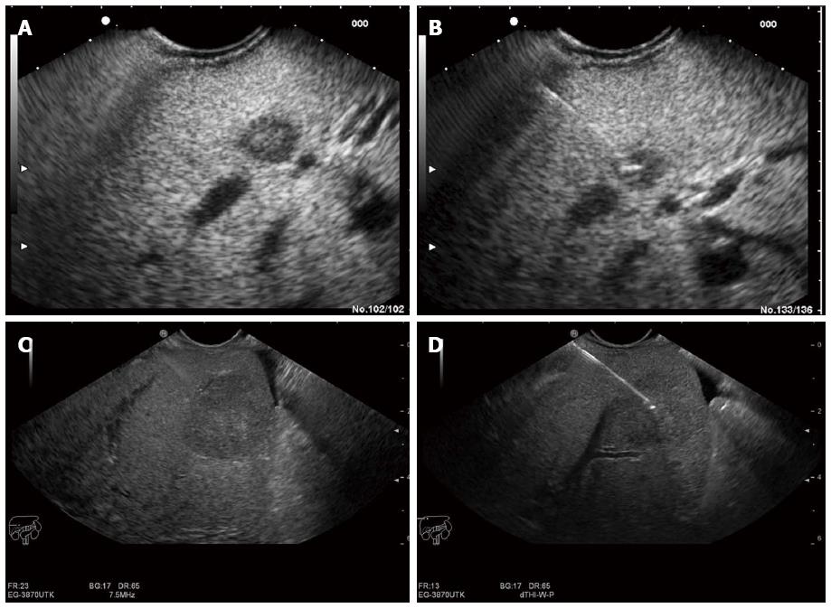
Figure 5 Endoscopic ultrasound image of lesion in the liver.
A: Endoscopic ultrasound (EUS) image of an 8 mm metastatic lesion in the liver; B: Endoscopic ultrasound (guided biopsy from the same lesion; C: EUS image of a 25 mm lesion in the liver; D: EUS guided aspiration biopsy from the same lesion.
- Citation: Srinivasan I, Tang SJ, Vilmann AS, Menachery J, Vilmann P. Hepatic applications of endoscopic ultrasound: Current status and future directions. World J Gastroenterol 2015; 21(44): 12544-12557
- URL: https://www.wjgnet.com/1007-9327/full/v21/i44/12544.htm
- DOI: https://dx.doi.org/10.3748/wjg.v21.i44.12544


