Copyright
©The Author(s) 2015.
World J Gastroenterol. Oct 28, 2015; 21(40): 11411-11427
Published online Oct 28, 2015. doi: 10.3748/wjg.v21.i40.11411
Published online Oct 28, 2015. doi: 10.3748/wjg.v21.i40.11411
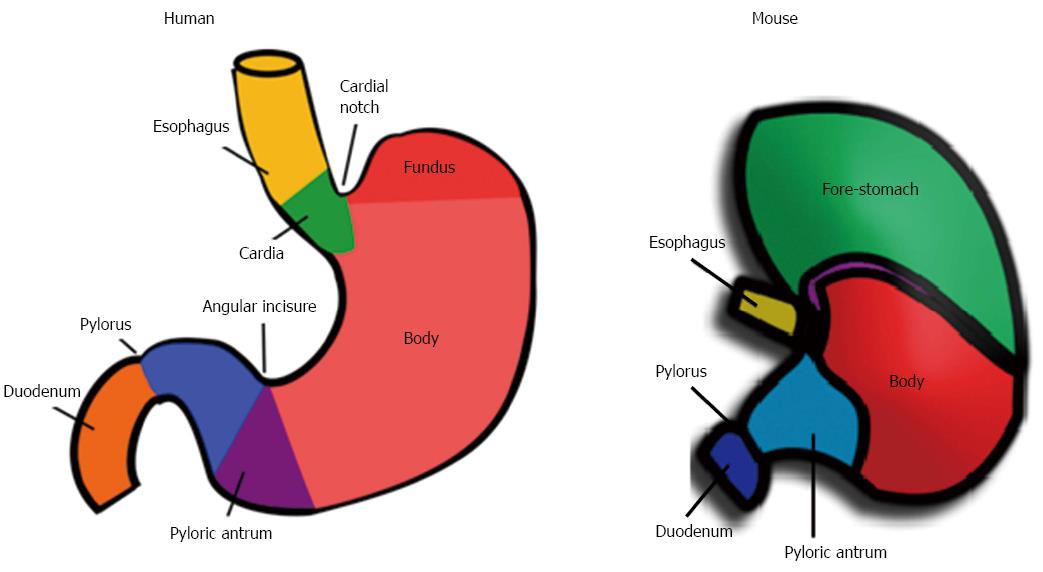
Figure 1 Gross anatomical characteristics of the human and mouse stomach.
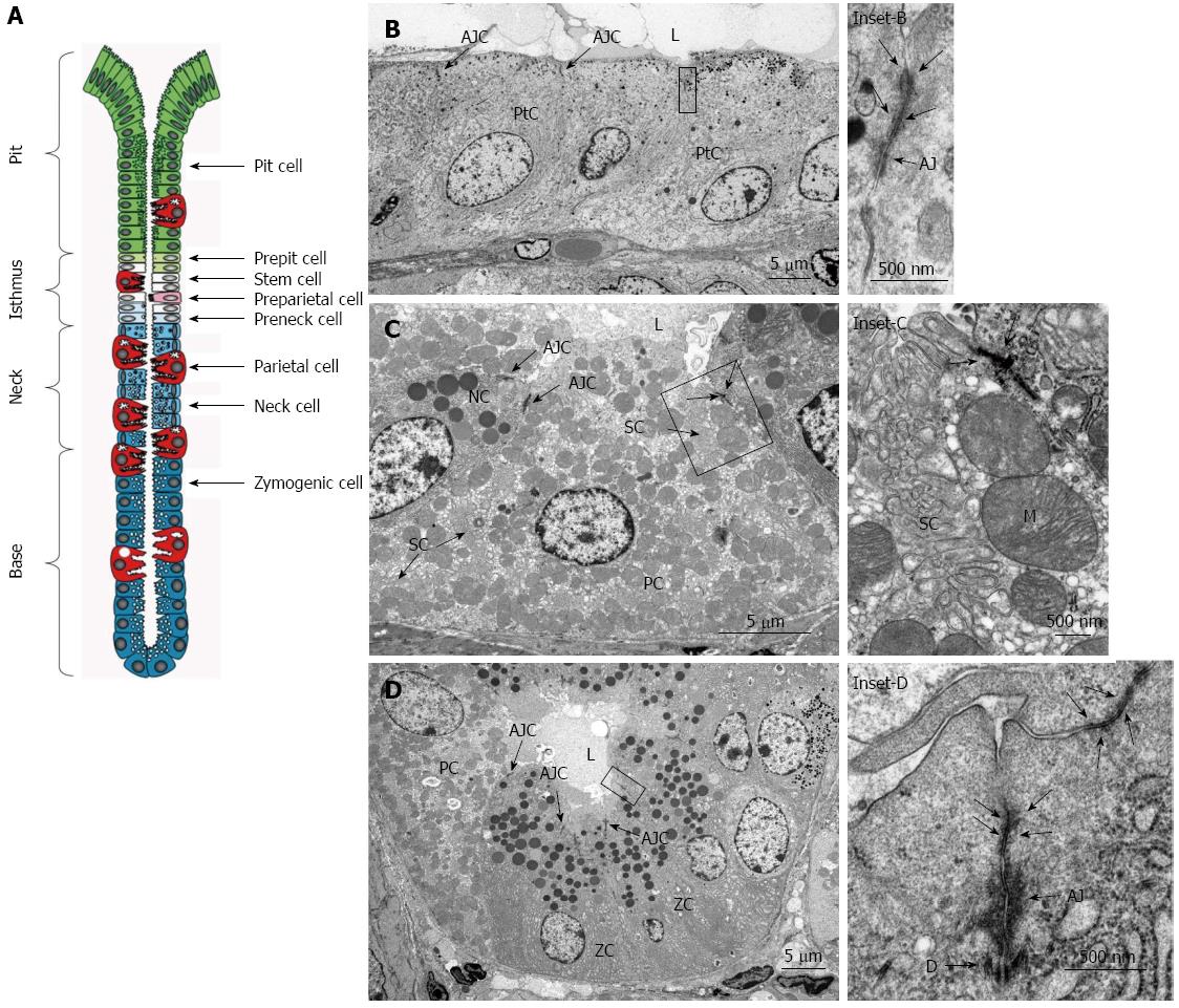
Figure 2 Histological structure of a gastric unit.
A: Diagrammatic representation of the organization of a gastric unit (also called a gastric gland), which contains a pit, isthmus, neck, and base. The location-specific cell types are identified in each region. Reproduced with permission from Karam SM[109]; B: Pit region cells (PtC) have apical junctional complexes (AJC) that are near the gastric lumen (L). Inset from the box in B: contains a tight junction (arrows) and an adherens junction (AJ). The desmosome is out of plane in this image. Bar in B is 5 μm and in the inset is 500 nm; C: Neck region cells, consisting mainly of parietal cells (PC) and neck cells (NC) also have AJC near the lumen of the gastric gland (L). Secretory canaliculi (SC) and mitochondria (M) are prominent in parietal cells. Inset from the box in C: contains a tight junction (arrows) and other parts of the apical junctional complex that are out of plane. Bar in C is 5 μm and in the inset is 500 nm; D: Base region cells consist mostly of zymogenic cells (ZCs) and a few PCs that also have AJC. Note that similar to the diagram in A, the apical cell cytoplasm of the zymogenic cells extends as a triangular wedge into the gland lumen (L) and apical junctional complexes are found at the lateral membranes where cells meet. Inset from the box in D: contains an apical junctional complex consisting of the tight junction (arrows), AJ, and desmome (D). Bar in D is 5 μm and in the inset is 500 nm.
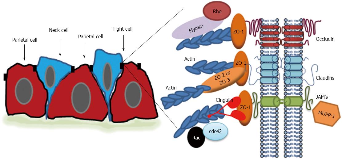
Figure 3 Schematic representation of the tight junction in gastric epithelial cells.
In the neck region of gastric glands, neck and parietal cells are oddly shaped but make tight junctions at the apical border between cells. Expanded diagram: Tight junctions in the stomach have classical components consisting of transmembrane proteins including occludin, claudins, and JAM proteins; peripheral scaffolding proteins like zonula occludens (ZO)-1, 2, and 3; linker proteins to the actin cytoskeleton like non-muscle myosin and cingulin; and signaling molecules like Rho, Rac, and cdc42. Actin filaments are also prominent.
-
Citation: Caron TJ, Scott KE, Fox JG, Hagen SJ. Tight junction disruption:
Helicobacter pylori and dysregulation of the gastric mucosal barrier. World J Gastroenterol 2015; 21(40): 11411-11427 - URL: https://www.wjgnet.com/1007-9327/full/v21/i40/11411.htm
- DOI: https://dx.doi.org/10.3748/wjg.v21.i40.11411


