Copyright
©The Author(s) 2015.
World J Gastroenterol. Oct 28, 2015; 21(40): 11260-11272
Published online Oct 28, 2015. doi: 10.3748/wjg.v21.i40.11260
Published online Oct 28, 2015. doi: 10.3748/wjg.v21.i40.11260
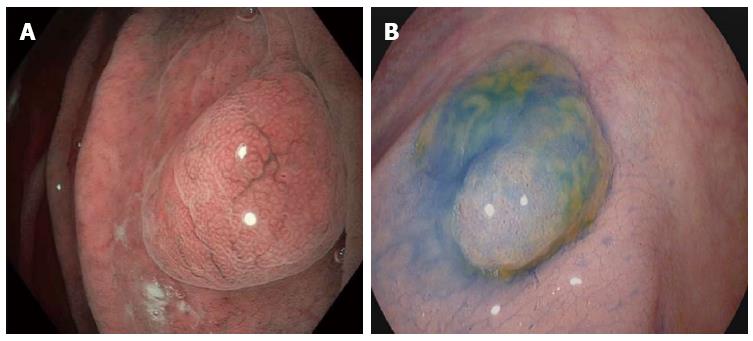
Figure 1 Dye-based chromoendoscopy and optical chromoendoscopy in the gastrointestinal tract.
Left picture: Optical dye-less chromoendoscopy with narrow band imaging (NBI) is based on the utilization of optical filters within the light source of the endoscope to narrow the bandwidth of spectral transmittance, thereby enhancing and facilitating the visualization of blood vessels. As exemplified on a fundic gland polyp in the stomach, NBI allows a clear delineation of the mucosal surface pit pattern architecture. Right picture: Dye-based chromoendoscopy with indigo carmine. Indigo carmine is a blue contrast agent that is used primarily in the colon for enhancing the detection or differentiation of colorectal neoplasms. As shown for a small colon polyp here, application of indigo carmine via a spraying catheter enhances the contrast and allows to visualize the pit pattern and to delineate mucosal irregularities.
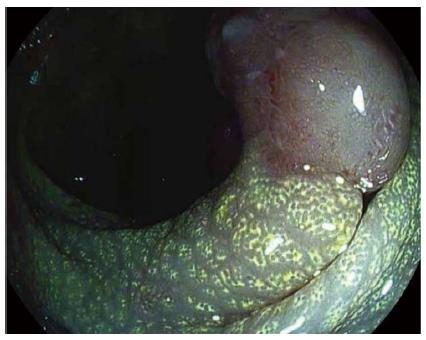
Figure 2 Digital dye-less chromoendocopy with i-scan in the lower gastrointestinal tract.
i-scan uses a digital postprocessing algorithm that reconstructs the endoscopic image from the video processor in real time resulting in improved contrast of the capillary patterns and enhancement of the mucosal surface pattern morphology as exemplified on a adenomatous polyp in the colon. As a result of an accumulation of lipid-filled macrophages within the lamina propria, the mucosa adjacent to the polyp exhibits a chicken skin mucosa on digital chromoendoscopy.
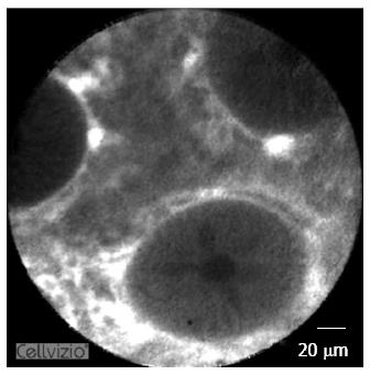
Figure 3 Confocal laser endomicroscopy in the lower gastrointestinal tract.
Confocal laser endomicroscopy (CLE) is based on the emission of a low power blue laser into the tissue after topical or systemic administration of contrast agents. The emitted light is then reflected from the tissue and refocused on the detection system by the same lens, leading to microscopic imaging at 1000-fold magnification in real time. As shown in healthy colonic mucosa in this picture, CLE allows a clear visualization of the colonic crypt architecture, single cells within the lamina propria and the microvasculature within the colon.
- Citation: Rath T, Tontini GE, Neurath MF, Neumann H. From the surface to the single cell: Novel endoscopic approaches in inflammatory bowel disease. World J Gastroenterol 2015; 21(40): 11260-11272
- URL: https://www.wjgnet.com/1007-9327/full/v21/i40/11260.htm
- DOI: https://dx.doi.org/10.3748/wjg.v21.i40.11260


