Copyright
©The Author(s) 2015.
World J Gastroenterol. Jan 28, 2015; 21(4): 1268-1274
Published online Jan 28, 2015. doi: 10.3748/wjg.v21.i4.1268
Published online Jan 28, 2015. doi: 10.3748/wjg.v21.i4.1268
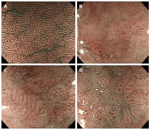
Figure 1 Representative endoscopic images of the microvascular pattern of depressed-type early gastric cancer, obtained using magnifying endoscopy combined with narrow-band imaging.
A: Normal; B: Irregular pattern, C: Regular pattern, D: Absent.
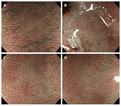
Figure 2 Representative endoscopic images of the microvascular pattern of depressed-type early gastric cancer, obtained using magnifying endoscopy combined with narrow-band imaging.
A: Normal; B: Fine-network pattern; C: Corkscrew pattern; D: Unclassified pattern.
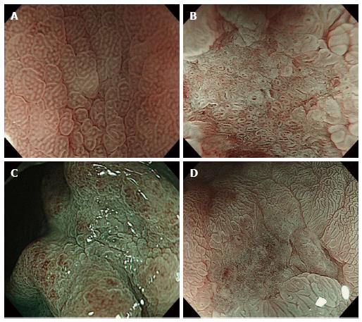
Figure 3 Representative endoscopic images of the mucosal surface pattern of depressed-type early gastric cancer, obtained with enhanced magnifying endoscopy using acetic acid staining.
A: Normal; B: Sessile barnacle pattern; C: Villous pattern; D: Unclassified pattern.
- Citation: Matsuo K, Takedatsu H, Mukasa M, Sumie H, Yoshida H, Watanabe Y, Akiba J, Nakahara K, Tsuruta O, Torimura T. Diagnosis of early gastric cancer using narrow band imaging and acetic acid. World J Gastroenterol 2015; 21(4): 1268-1274
- URL: https://www.wjgnet.com/1007-9327/full/v21/i4/1268.htm
- DOI: https://dx.doi.org/10.3748/wjg.v21.i4.1268


