Copyright
©The Author(s) 2015.
World J Gastroenterol. Jan 28, 2015; 21(4): 1125-1139
Published online Jan 28, 2015. doi: 10.3748/wjg.v21.i4.1125
Published online Jan 28, 2015. doi: 10.3748/wjg.v21.i4.1125
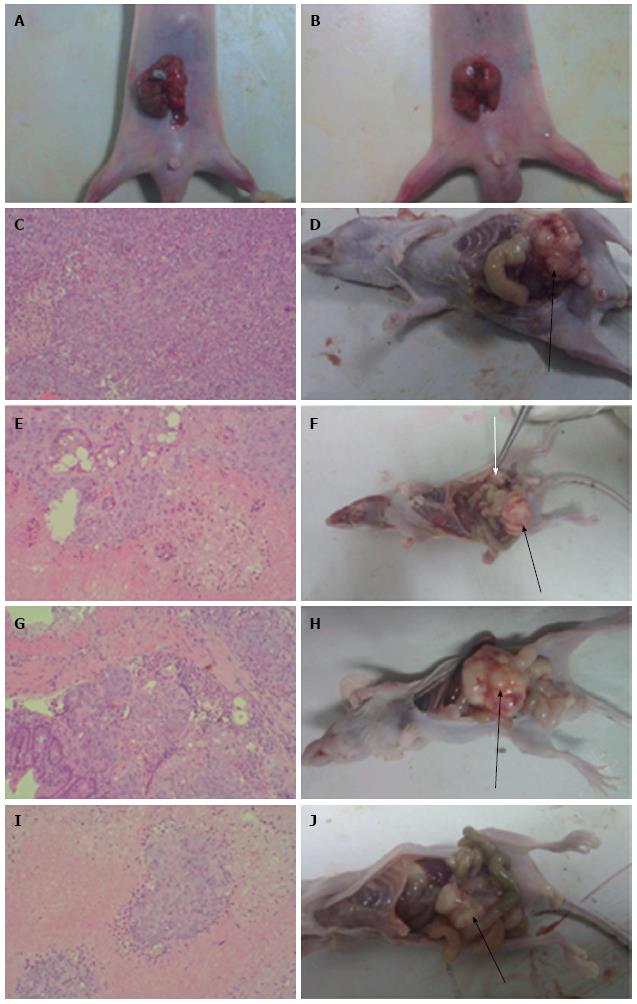
Figure 1 Pathology of primary colon cancer tissues (HE staining) and gross anatomy of nude mice implanted with human colon cancer HCT-116 cells.
A, B: Tumor pieces were transplanted into the cecum of nude mice by purse-string suture; C, E, G and I: HE staining of colon cancer tissues from mice in the CON, WCA, 5-FU and WCA + 5-FU groups (magnification × 100); D, F, H and J: Gross anatomy of mice in the CON, WCA, 5-FU and WCA + 5-FU groups. Black arrows indicate orthotopic tumors, and white arrows indicate the abdominal wall tumor. WCA: Weichang’an; 5-FU: 5-fluorouracil; CON: Control.
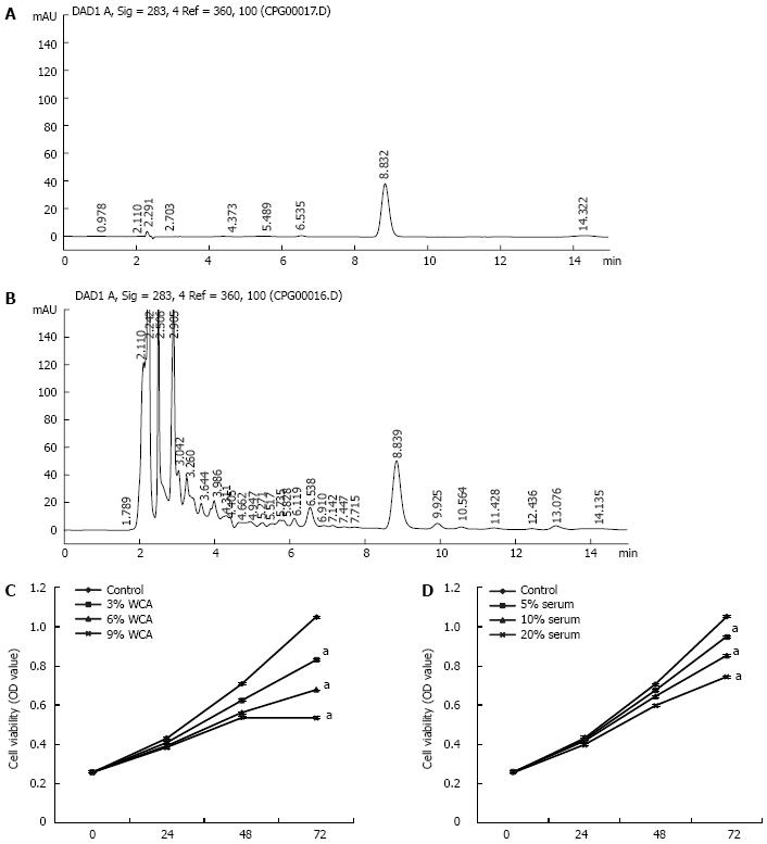
Figure 2 HPLC fingerprinting of hesperidin (A) and Weichang’an (B); HCT-116 cells were treated with different concentrations of Weichang’an (C) and different concentrations of Weichang’an-containing serum (D) in vitro.
Proliferation of cells was detected using the MTT assay. Cell viability was expressed as optical density (OD) value (mean ± SD). aP < 0.05 vs cell viability of the control group at the same time point. WCA: Weichang’an.
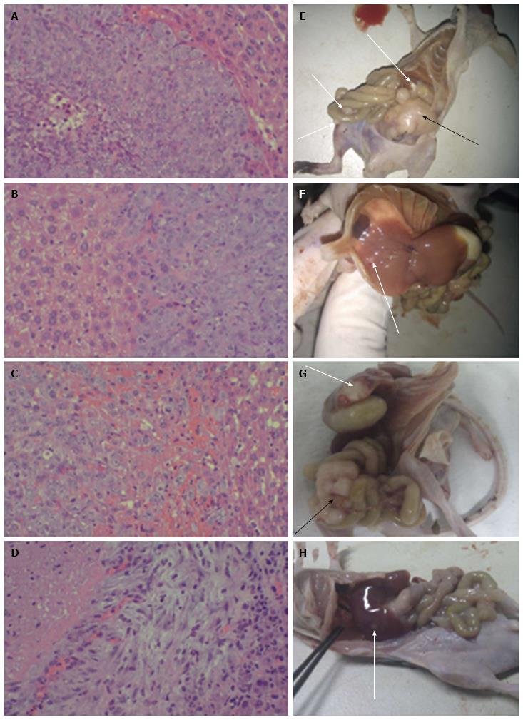
Figure 3 Hepatic metastases of colon cancer in the orthotopic transplant nude mouse model.
A-D: HE staining of hepatic metastases tissues from mice in the CON, WCA, 5-FU and WCA + 5-FU groups (magnification × 200); E-H: Gross anatomy of mice in the CON, WCA, 5-FU and WCA + 5-FU groups. The black arrow indicates orthotopic tumor, the long white arrow indicates the hepatic metastatic tumor, and the short white arrow indicates the metastasis of colon cancer to the intestinal wall. WCA: Weichang’an; 5-FU: 5-fluorouracil; CON: Control.
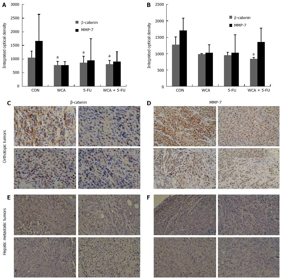
Figure 4 Immunohistochemistry detection of β-catenin and matrix metalloproteinase-7 in orthotopic tumors and hepatic metastatic tumors.
A, B: Integrated optical density (OD) of β-catenin and matrix metalloproteinase (MMP)-7 protein expressed in orthotopic tumors (A) or hepatic metastatic tumors (B) from mice in the CON, WCA, 5-FU and WCA + 5-FU groups. Data are presented as mean ± SD and aP < 0.05 vs corresponding CON group; C: IHC staining for β-catenin in orthotopic tumors (magnification × 200); D: IHC staining for MMP-7 in orthotopic tumors (magnification × 200); E: IHC staining for β-catenin in hepatic metastatic tumors (magnification × 200); F: IHC staining for MMP-7 in hepatic metastatic tumors (magnification × 200). WCA: Weichang’an; 5-FU: 5-fluorouracil; CON: Control.
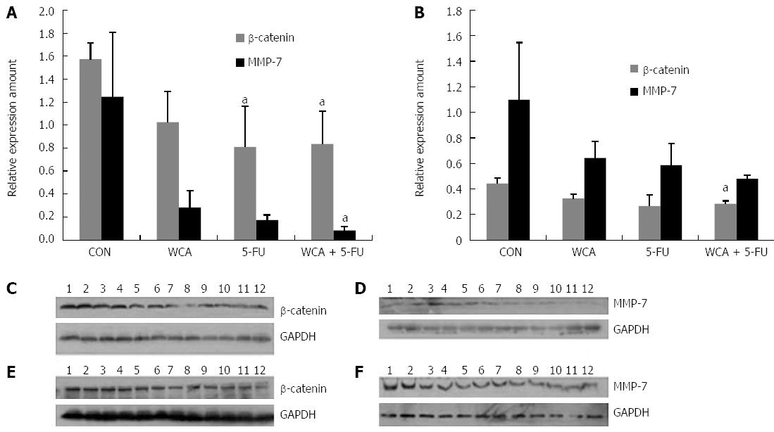
Figure 5 Western blot analysis of β-catenin and matrix metalloproteinase-7 protein expression in orthotopic and hepatic metastatic tumors.
A, B: Quantification of β-catenin and matrix metalloproteinase (MMP)-7 expression in orthotopic tumors (A) and hepatic metastatic tumors (B). Data were presented as mean ± SD and aP < 0.05 vs corresponding CON group; C: β-catenin expression in orthotopic tumors; D: MMP-7 expression in orthotopic tumors; E: β-catenin expression in hepatic metastatic tumors; F: MMP-7 expression in hepatic metastatic tumors. Lanes 1-3, CON group; 4-6, WCA group; 7-9, 5-FU group; and 10-12, WCA + 5-FU group. WCA: Weichang’an; 5-FU: 5-fluorouracil; CON: Control.
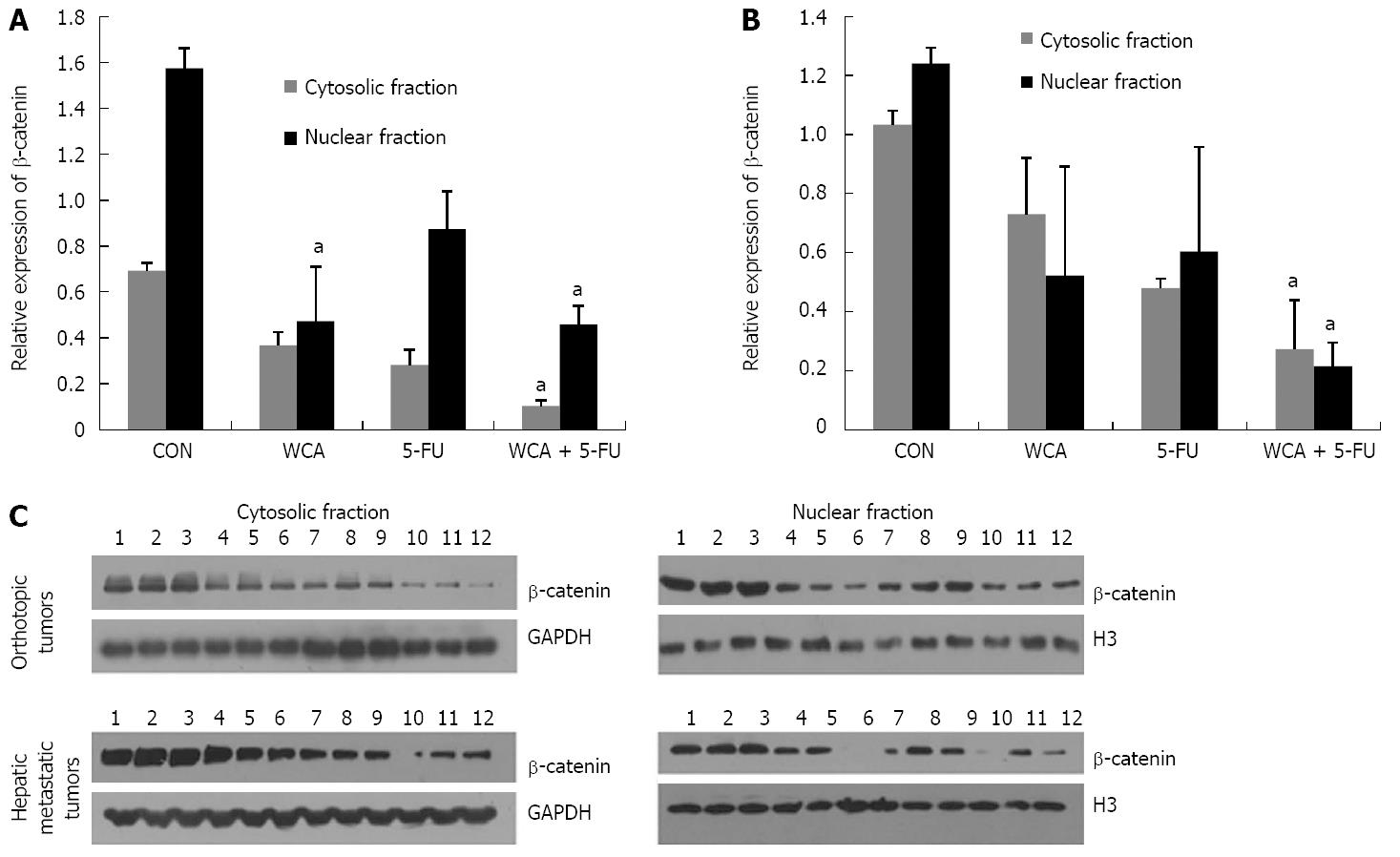
Figure 6 Cell fractionation and western blot analysis of β-catenin protein expression in both orthotopic tumors and hepatic metastatic tumors.
A, B: Quantification of β-catenin expression in cytosolic fraction and nuclear fraction in orthotopic tumors (A) and in hepatic metastatic tumors (B). Data were presented as mean ± SD and aP < 0.05 vs corresponding CON group; C: β-catenin expression in cytosolic fraction and nuclear fraction in orthotopic tumors and hepatic metastatic tumors. Lanes 1-3, CON group; 4-6, WCA group; 7-9, 5-FU group; and 10-12, WCA + 5-FU group. WCA: Weichang’an; 5-FU: 5-fluorouracil; CON: Control.
- Citation: Tao L, Yang JK, Gu Y, Zhou X, Zhao AG, Zheng J, Zhu YJ. Weichang’an and 5-fluorouracil suppresses colorectal cancer in a mouse model. World J Gastroenterol 2015; 21(4): 1125-1139
- URL: https://www.wjgnet.com/1007-9327/full/v21/i4/1125.htm
- DOI: https://dx.doi.org/10.3748/wjg.v21.i4.1125


