Copyright
©The Author(s) 2015.
World J Gastroenterol. Jan 28, 2015; 21(4): 1091-1098
Published online Jan 28, 2015. doi: 10.3748/wjg.v21.i4.1091
Published online Jan 28, 2015. doi: 10.3748/wjg.v21.i4.1091
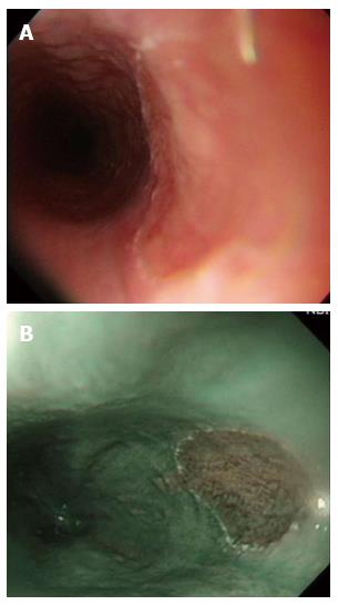
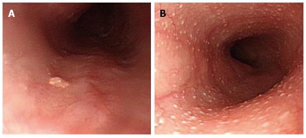
Figure 1 Heterotopic gastric mucosa.
A: Heterotopic gastric mucosa appears salmon-colored under conventional endoscopy and is recognized as flat or slightly elevated; B: Narrow band imaging facilitates mucosal surface evaluation of heterotopic gastric mucosa by adjusting reflected light to enhance the contrast between the esophageal mucosa and the gastric mucosa and may improve the diagnosis of heterotopic gastric mucosa.
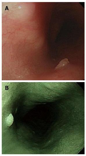
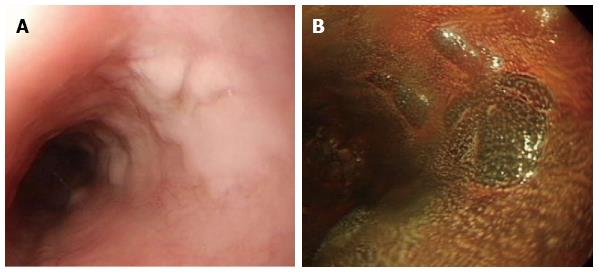
Figure 2 Squamous cell papilloma.
A: Squamous cell papilloma is recognized as whitish-pink, wart-like exophytic projections on conventional endoscopy; B: Narrow band imaging facilitates mucosal surface evaluation of squamous cell papilloma and shows that microvessels in the lesion are not dilated.
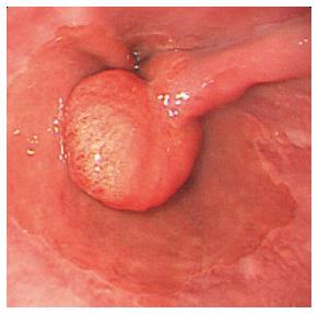
Figure 3 Hyperplasia polyp.
Hyperplasia polyp occurs as a polypoid lesion and is located on edematous inflamed gastric folds at the gastroesophageal junction.
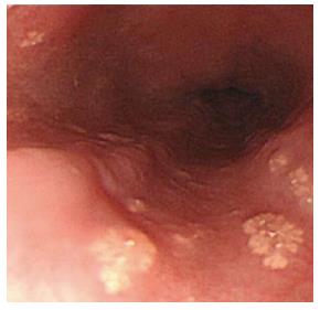
Figure 4 Xanthoma.
Xanthomas are endoscopically recognized as elevated, granular, yellowish, fern-like lesions and scattered on a normal mucosal surface.
Figure 5 Ectopic sebaceous gland.
A: The number of ectopic sebaceous gland is variable from single to B: more than one hundred yellowish plaques measuring 1 to 2 mm in the esophagus.
Figure 6 Glycogenic acanthosis.
A: Esophagogastroduodenoscopy reveals multiple, uniformly sized, oval or round glycogenic acanthosis usually < 1 cm, involving otherwise normal esophageal mucosa; B: In chromoscopy with iodine spray, glycogenic acanthosis is recognized as slightly elevated iodine-positive, brownish areas.
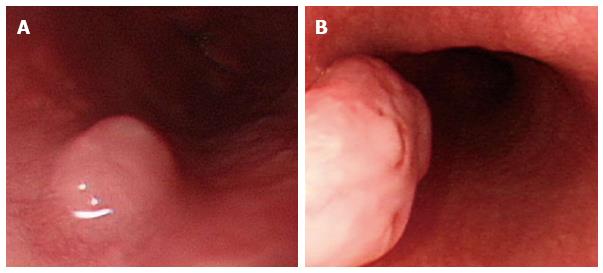
Figure 7 Leiomyoma.
A: Leiomyoma commonly arises from the muscularis propria layer of the esophagus and presents as submucosal tumor; B: Leiomyoma arising from the muscularis mucosae can present as a polypoid intraluminal tumor.
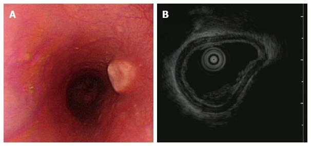
Figure 8 Granular cell tumor.
A: Granular cell tumor is endoscopically recognized as a firm, yellowish subepithelial tumor covered with the normal mucosa in the esophagus; B: Endoscopic ultrasonography of granular cell tumor shows a homogenous hypo-echogenic tumor extending from the muscularis mucosa layer, and the musculais propria is not involved.
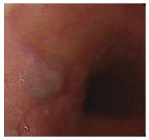
Figure 9 Hemangioma.
On endoscopy, esophageal hemangioma appears cystic and bluish-red and can be pressed with biopsy forceps.
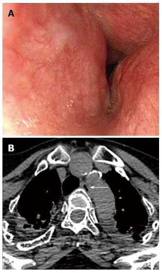
Figure 10 Dysphagia aortica.
A: Esophagogastroduodenoscopy reveals a pulsatile extrinsic compression at about 25 cm from the incisor; B: The chest computed tomography showed aortic arch and descending aorta tortuosity with compression into adjacent esophagus. The arrow indicates the esophagus.
- Citation: Tsai SJ, Lin CC, Chang CW, Hung CY, Shieh TY, Wang HY, Shih SC, Chen MJ. Benign esophageal lesions: Endoscopic and pathologic features. World J Gastroenterol 2015; 21(4): 1091-1098
- URL: https://www.wjgnet.com/1007-9327/full/v21/i4/1091.htm
- DOI: https://dx.doi.org/10.3748/wjg.v21.i4.1091


