Copyright
©The Author(s) 2015.
World J Gastroenterol. Sep 28, 2015; 21(36): 10468-10474
Published online Sep 28, 2015. doi: 10.3748/wjg.v21.i36.10468
Published online Sep 28, 2015. doi: 10.3748/wjg.v21.i36.10468
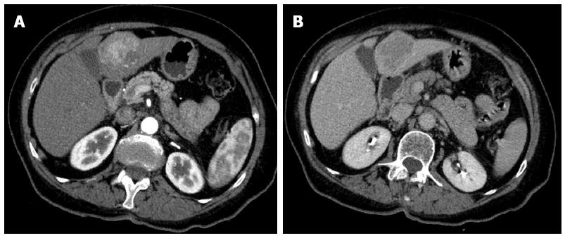
Figure 1 Computerized tomography scan.
Left liver neoplasm (segments III and IV), hypervascular (A) with rapid portal-phase wash-out (B).
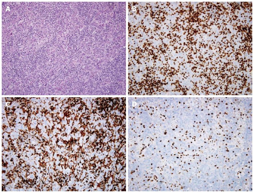
Figure 2 Neoplastic cells surrounded by a dense lymphoid infiltrate.
A: Hematoxylin/eosin stain x 200; B: CD3 immunostain x 200; C: CD4 immunostain x 200; D: CD8 immunostain x 200.
- Citation: Insilla AC, Faviana P, Pollina LE, Simone PD, Coletti L, Filipponi F, Campani D. Lymphoepithelioma-like hepatocellular carcinoma: Case report and review of the literature. World J Gastroenterol 2015; 21(36): 10468-10474
- URL: https://www.wjgnet.com/1007-9327/full/v21/i36/10468.htm
- DOI: https://dx.doi.org/10.3748/wjg.v21.i36.10468


