Copyright
©The Author(s) 2015.
World J Gastroenterol. Sep 28, 2015; 21(36): 10461-10467
Published online Sep 28, 2015. doi: 10.3748/wjg.v21.i36.10461
Published online Sep 28, 2015. doi: 10.3748/wjg.v21.i36.10461
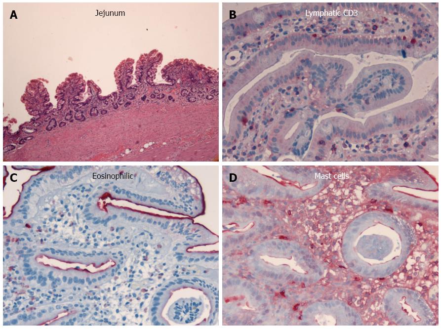
Figure 1 Histopathologic diagnosis of chronic malabsorption syndrome associated with exudative enteropathy as a result of prolonged intestinal primary lymphangectasia and chronic jejunitis of undefined etiology.
A: The jejunum showed slight architectural deviations with flat mucosa due to partial villous atrophy; B: T cell lymphatic infiltrates were normal as seen in CD3 staining, which ruled out celiac disease and lymphoma; The amounts of C: Eosinophilic granulocytes; and D: Mast cells were normal, thus excluding mastocytosis and eosinophilic gastroenteritis. The histological sections were prepared with hematoxylin-eosin staining (magnification × 40) and additional immunohistochemistry was performed.
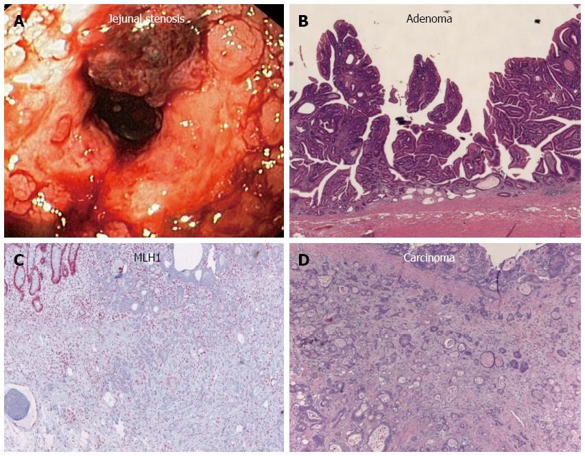
Figure 2 Disease progression.
A: Push enteroscopy and high-pressure balloon dilatation of the stenosis were performed; Histology indicated the development of an B: Adenoma; and D: Carcinoma. Thus, 20 years of persistent inflammation led to malignancy according to an enteritis-dysplasia-adenocarcinoma sequence; C: Unaffected crypts showed strong nuclear MLH1 expression, while neoplastic tissue failed to express MLH1. Histological sections A-D were prepared with hematoxylin-eosin staining (magnification × 40) and additional immunohistochemistry was performed.
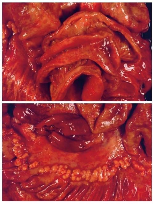
Figure 3 Postmortem confirmation of brown bowel syndrome associated with multifocal MLH1-positive small bowel adenocarcinoma.
- Citation: Raithel M, Rau TT, Hagel AF, Albrecht H, Rossi T, Kirchner T, Hahn EG. Jejunitis and brown bowel syndrome with multifocal carcinogenesis of the small bowel. World J Gastroenterol 2015; 21(36): 10461-10467
- URL: https://www.wjgnet.com/1007-9327/full/v21/i36/10461.htm
- DOI: https://dx.doi.org/10.3748/wjg.v21.i36.10461


