Copyright
©The Author(s) 2015.
World J Gastroenterol. Aug 28, 2015; 21(32): 9503-9511
Published online Aug 28, 2015. doi: 10.3748/wjg.v21.i32.9503
Published online Aug 28, 2015. doi: 10.3748/wjg.v21.i32.9503
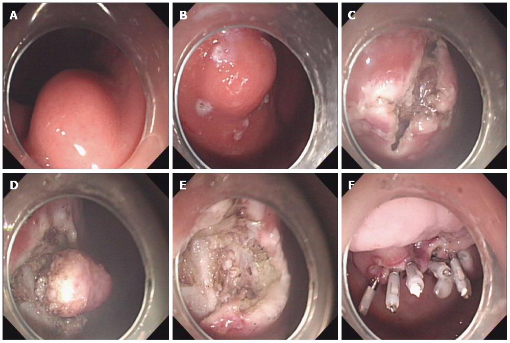
Figure 1 Endoscopic muscularis excavation.
A: A subepithelial tumor was found at the posterior wall of the gastric body; B: Making several dots around the tumor; C: A cross-incision was made at the overlying mucosa of the tumor; D: Excavating the tumor from the muscularis propria layer; E: An artificial ulcer was observed after excavation; F: The artificial ulcer was closed with several clips.
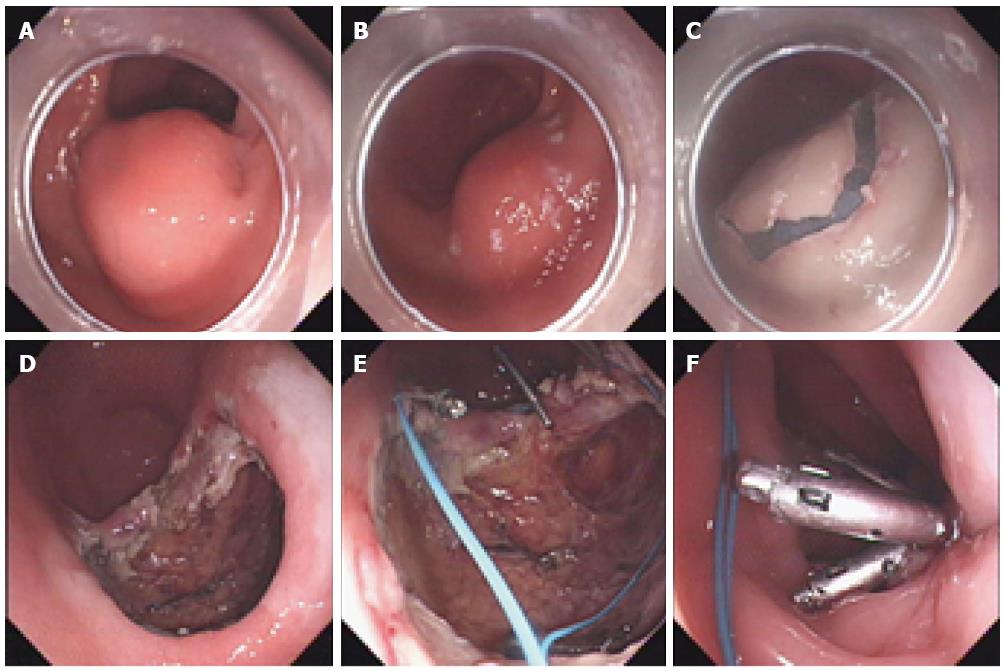
Figure 2 Endoscopic full-thickness resection.
A: A subepithelial tumor was found at the greater curvature of the gastric antrum; B: Making several dots around the tumor; C: The mucosa incision was incised outside the marker dots; D: The omentum could be seen through the gastric wall defect after endoscopic full-thickness resection; E, F: Closure of the gastric wall defect using clips and an endoloop.
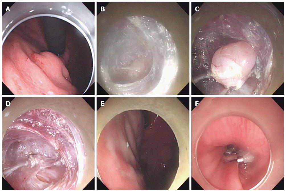
Figure 3 Submucosal tunneling endoscopic resection.
A: A subepithelial tumor was found in the gastric fundus; B, C: Creating a submucosal tunnel to the tumor and then excavating the tumor from the muscularis propria layer via the submucosal tunnel; D: The omentum could be seen through a small gastric wall after tumor removal; E: The gastric mucosa was intact; F: The entrance of the submucosal tunnel was closed with several clips after tumor removal.
- Citation: Zhang Y, Ye LP, Mao XL. Endoscopic treatments for small gastric subepithelial tumors originating from muscularis propria layer. World J Gastroenterol 2015; 21(32): 9503-9511
- URL: https://www.wjgnet.com/1007-9327/full/v21/i32/9503.htm
- DOI: https://dx.doi.org/10.3748/wjg.v21.i32.9503


