Copyright
©The Author(s) 2015.
World J Gastroenterol. Jan 21, 2015; 21(3): 997-1000
Published online Jan 21, 2015. doi: 10.3748/wjg.v21.i3.997
Published online Jan 21, 2015. doi: 10.3748/wjg.v21.i3.997
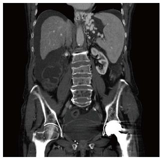
Figure 1 Oesophageal and gastric varices.
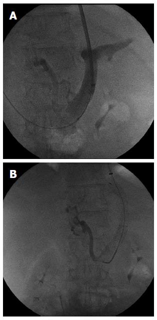
Figure 2 Portography.
A: Transjugular intrahepatic porto-systemic shunt 1; B: Transjugular intrahepatic porto-systemic shunt 2.
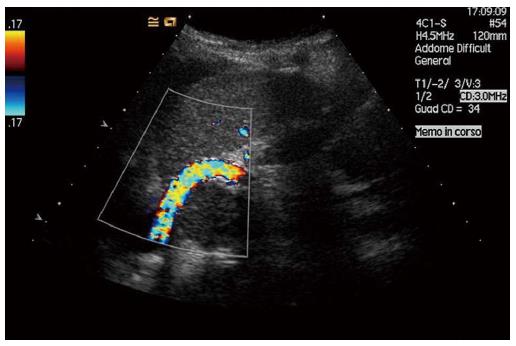
Figure 3 Ecocolor Doppler transjugular intrahepatic porto-systemic shunt.
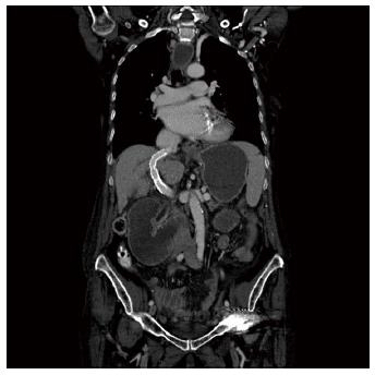
Figure 4 Computed tomography.
A: Computed tomography after a transjugular intrahepatic porto-systemic shunt; B: Coil embolization of right gastric vein.
- Citation: Liverani A, Solinas L, Cesare TD, Velari L, Neri T, Cilurso F, Favi F, Bizzarri G. Preoperative trans-jugular porto-systemic shunt for oncological gastric surgery in a cirrhotic patient. World J Gastroenterol 2015; 21(3): 997-1000
- URL: https://www.wjgnet.com/1007-9327/full/v21/i3/997.htm
- DOI: https://dx.doi.org/10.3748/wjg.v21.i3.997


