Copyright
©The Author(s) 2015.
World J Gastroenterol. Jan 21, 2015; 21(3): 969-976
Published online Jan 21, 2015. doi: 10.3748/wjg.v21.i3.969
Published online Jan 21, 2015. doi: 10.3748/wjg.v21.i3.969
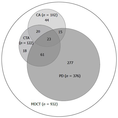
Figure 1 Patients included (grey) in the analysis and those excluded (white).
PD: Pancreaticoduodenectomy; CTA: Computed tomography angiography; CA: Conventional angiography; MDCT: Multidetector computed tomography.
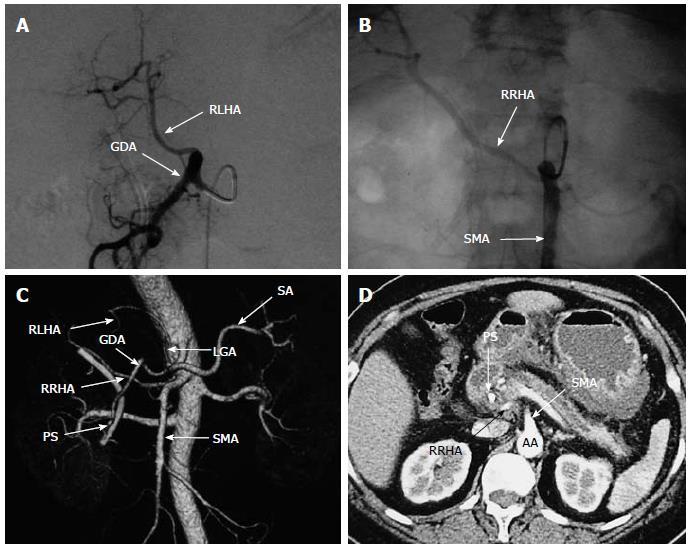
Figure 2 Sixty-year-old woman with replaced right hepatic artery and replaced left hepatic artery.
A: Visceral angiogram with celiac injection demonstrating an RLHA; B: Visceral angiogram with SMA injection revealed an RRHA originating from the proximal SMA; C: Volume-rendered reformatted image depicting Michels type IV; D: MDCT showing an RRHA originating from the SMA traveling behind the portal vein. RRHA: Replaced right hepatic artery; RLHA: Replaced left hepatic artery; SMA: Superior mesenteric artery; SA: Splenic artery; GDA: Gastroduodenal artery; LGA: Left gastric artery; PS: Plastic stent; AA: Abdominal aorta.
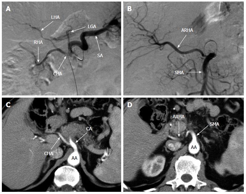
Figure 3 Sixty-five-year-old man with an accessory right hepatic artery.
Conventional angiography with celiac injection (A) demonstrating a classic hepatic arterial anatomy, and one with SMA injection (B) showing an ARHA originating from the proximal SMA. MDCT showing the CHA arising from the CA (C), and an ARHA originating from the SMA, traveling behind the pancreatic head (D). ARHA: Accessory right hepatic artery; CHA: Common hepatic artery; SA: Splenic artery; LGA: Left gastric artery; RHA: Right hepatic artery; LHA: Left hepatic artery; SMA: Superior mesenteric artery; AA: Abdominal aorta; CA: Celiac axis.
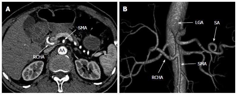
Figure 4 Sixty-eight-year-old man with a replaced common hepatic artery.
A: MDCT demonstrating an RCHA originating from the SMA, running behind the superior mesenteric vein, with no common hepatic artery found arising from the celiac axis; B: Volumetric three-dimensional CT angiography demonstrating Michels type IX. RCHA: Replaced common hepatic artery; MDCT: Multidetector computed tomography; CT: Computed tomography; SMA: Superior mesenteric artery; SA: Splenic artery; LGA: Left gastric artery; AA: Abdominal aorta.
Figure 5 Sixty-five-year-old man with celiac axis stenosis.
Computed tomography scan of the abdomen (A) and celiac artery angiogram (B) revealing stenosis of the celiac axis (arrow). (C) Superior mesenteric arteriogram demonstrating retrograde filling of the hepatic artery through a dilatation of the pancreaticoduodenal arcade.
- Citation: Yang F, Di Y, Li J, Wang XY, Yao L, Hao SJ, Jiang YJ, Jin C, Fu DL. Accuracy of routine multidetector computed tomography to identify arterial variants in patients scheduled for pancreaticoduodenectomy. World J Gastroenterol 2015; 21(3): 969-976
- URL: https://www.wjgnet.com/1007-9327/full/v21/i3/969.htm
- DOI: https://dx.doi.org/10.3748/wjg.v21.i3.969


