Copyright
©The Author(s) 2015.
World J Gastroenterol. Jan 21, 2015; 21(3): 935-943
Published online Jan 21, 2015. doi: 10.3748/wjg.v21.i3.935
Published online Jan 21, 2015. doi: 10.3748/wjg.v21.i3.935
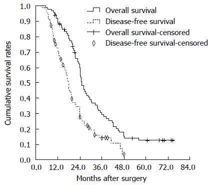
Figure 1 Overall survival and disease-free survival curves of the 85 hepatitis B virus-associated intrahepatic cholangiocarcinoma patients after curative resection.
The median overall survival was 25 mo. The median disease-free survival was 17.9 mo.
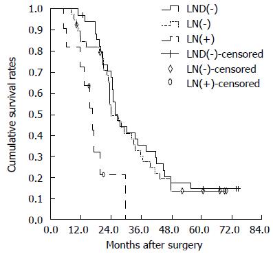
Figure 2 Overall survival curves of patients in lymph node dissection (-), lymph node (-) and lymph node (+) groups.
There was no statistically significant difference in OS between the lymph node dissection (LND) (-) and LN (-) groups (P = 0.729). There were significant differences between LN (-) and LN (+) groups (P = 0.033). A statistically significant difference between LND (-) and LN (+) groups was observed (P = 0.004). LND: Lymph node dissection; LN: Lymph node.
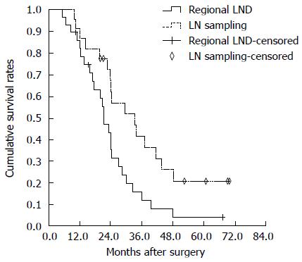
Figure 3 Overall survival curves of hepatitis B virus-associated intrahepatic cholangiocarcinoma patients undergoing sampling lymph node sampling and regional lymph node dissection.
There were no significant differences between the two groups (P = 0.089). LND: Lymph node dissection; LN: Lymph node.
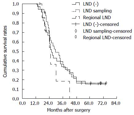
Figure 4 Overall survival curves of hepatitis B virus-associated intrahepatic cholangiocarcinoma patients without lymph node involvement.
There was no survival difference between lymph node sampling and regional LND groups (P = 0.182). There was no statistically significant difference in overall survival between LND (-) and lymph node sampling groups (P = 0.709). There were also no significant differences between LND (-) and regional LND groups (P = 0.309). LND: Lymph node dissection.
- Citation: Wu ZF, Wu XY, Zhu N, Xu Z, Li WS, Zhang HB, Yang N, Yao XQ, Liu FK, Yang GS. Prognosis after resection for hepatitis B virus-associated intrahepatic cholangiocarcinoma. World J Gastroenterol 2015; 21(3): 935-943
- URL: https://www.wjgnet.com/1007-9327/full/v21/i3/935.htm
- DOI: https://dx.doi.org/10.3748/wjg.v21.i3.935


