Copyright
©The Author(s) 2015.
World J Gastroenterol. Jan 21, 2015; 21(3): 820-828
Published online Jan 21, 2015. doi: 10.3748/wjg.v21.i3.820
Published online Jan 21, 2015. doi: 10.3748/wjg.v21.i3.820
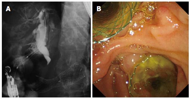
Figure 1 Endoscopic ultrasound-guided choledocoduodenostomy combined with duodenal stent.
A: Endoscopic ultrasound-guided choledocoduodenostomy (EUS-CDS) was performed from duodenal bulb after duodenal stenting; B: Endoscopic view of EUS-CDS combined with duodenal stent.
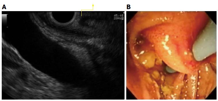
Figure 2 Double puncture of duodenal mucosa.
A: Double mucosa of duodenum on endoscopic ultrasound view (arrow); B: Endoscopic view of double puncture of duodenal mucosa.
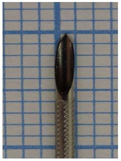
Figure 3 Novel endoscopic ultrasound-guided fine needle aspiration needle (Sono Tip Pro Control 19 G needle, Medi-Globe GmbH, Rosenheim, Germany).
The cut surface of this fine needle aspiration needle is 5 mm, and this needle has sharpness.
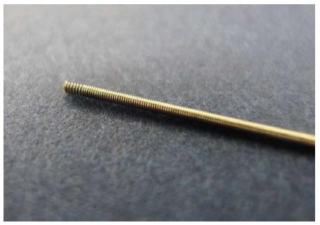
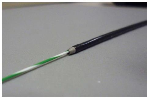
Figure 4 Cyst-wire (Medi-Globe GmbH, Rosenheim, Germany).
To avoid wire sharing, top formation of this guidewire is coil.
Figure 5 Cysto-Gastro-Set (Endoflex, GmbH, Voerde, Germany).
This devise is always coaxial with the guidewire.
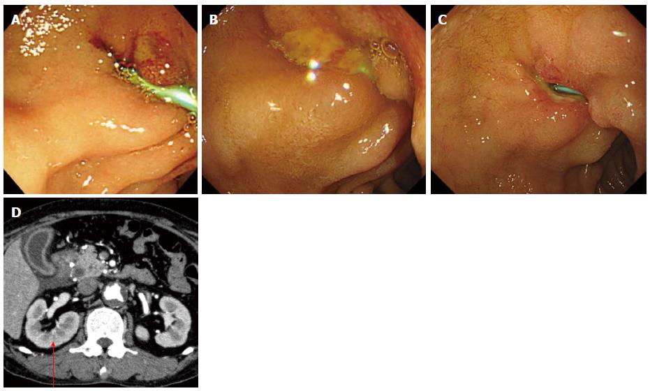
Figure 6 Bile leak due to plastic stent of endoscopic ultrasound-guided choledocoduodenostomy.
A: Endoscopic view of day 1; B: Day 2; C: Day 3; D: Computed tomography showed bile leak (arrow).
- Citation: Ogura T, Higuchi K. Technical tips of endoscopic ultrasound-guided choledochoduodenostomy. World J Gastroenterol 2015; 21(3): 820-828
- URL: https://www.wjgnet.com/1007-9327/full/v21/i3/820.htm
- DOI: https://dx.doi.org/10.3748/wjg.v21.i3.820


