Copyright
©The Author(s) 2015.
World J Gastroenterol. Jan 21, 2015; 21(3): 1044-1048
Published online Jan 21, 2015. doi: 10.3748/wjg.v21.i3.1044
Published online Jan 21, 2015. doi: 10.3748/wjg.v21.i3.1044
Figure 1 Colonoscopy and histologic findings in the early onset of colon inflammation.
A: Inflammation in the colon, no polyps were found in the colon; B: Mucosal epithelial necrosis, destruction, distortion and branching of lamina propria glands. The distance between the glands in some regions was increased and goblet cells decreased significantly; a large number of lymphocytes and plasma cells infiltrated the lamina propria, there were neutrophils in crypts, and lymphocytes increased significantly in the base of the mucosa; a large number of lymphocytes and plasma cells infiltrated the submucosa; C: Lamina propria glands were distorted and branched accompanied by crypt abscesses and parts of the glands showed atrophy. The distance between the glands in some regions was increased, in which goblet cells were decreased. Inflammation in the lamina propria was uniform, with infiltration of numerous lymphocytes and plasma cells, and stromal vessels were significantly dilated and congested (HE staining).
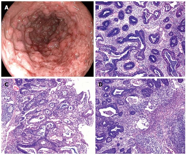
Figure 2 Colonoscopy and histologic finding 6 mo later.
A: Diffuse congestion and edema in the mucosa accompanied by focal hemorrhage and exudation, showing the different shapes, sizes, erosive lesions and superficial ulcers, and most of the regional mucosal hyperplasia was granular with pseudopolyp formation; B-D: Formation of necrosis and shallow ulcers in part of the mucosal epithelial and glands in the lamina propria were distorted and branching. Several glands showed atrophy and goblet cells were decreased. Neutrophils infiltrated the lamina propria. The stromal blood vessels were significantly dilated and congested (HE staining).
- Citation: Feng JS, Ye Y, Guo CC, Luo BT, Zheng XB. Ulcerative colitis with inflammatory polyposis in a teenage boy: A case report. World J Gastroenterol 2015; 21(3): 1044-1048
- URL: https://www.wjgnet.com/1007-9327/full/v21/i3/1044.htm
- DOI: https://dx.doi.org/10.3748/wjg.v21.i3.1044


