Copyright
©The Author(s) 2015.
World J Gastroenterol. Jan 21, 2015; 21(3): 1028-1031
Published online Jan 21, 2015. doi: 10.3748/wjg.v21.i3.1028
Published online Jan 21, 2015. doi: 10.3748/wjg.v21.i3.1028
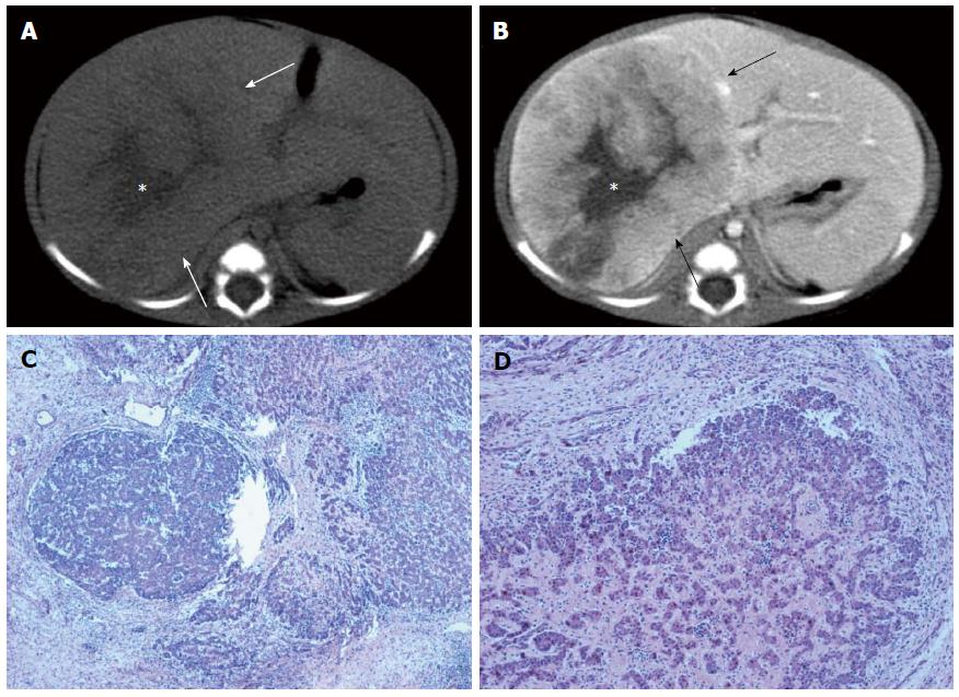
Figure 1 Computed tomography findings.
A: Non-contrast computed tomography (CT) of the liver showed a slightly hypo-dense mass in the right lobe of the liver (arrow) with a hypo-dense central star-like scar (asterisk); B: Contrast-enhanced CT of the liver in the portal venous phase showed inhomogeneous and intense enhancement of the mass (arrow), with the central star-like scar (asterisk); C: Histopathology showed typical focal nodular hyperplasia, with hepatocytic nodules separated by bands of fibrous tissue (HE stain, original magnification × 50); D: Foci of hepatoblastoma showed epithelioid tumor cells separated by proliferated fibrous tissue (HE stain, original magnification × 100).
- Citation: Gong Y, Chen L, Qiao ZW, Ma YY. Focal nodular hyperplasia coexistent with hepatoblastoma in a 36-d-old infant. World J Gastroenterol 2015; 21(3): 1028-1031
- URL: https://www.wjgnet.com/1007-9327/full/v21/i3/1028.htm
- DOI: https://dx.doi.org/10.3748/wjg.v21.i3.1028


