Copyright
©The Author(s) 2015.
World J Gastroenterol. Jul 28, 2015; 21(28): 8730-8738
Published online Jul 28, 2015. doi: 10.3748/wjg.v21.i28.8730
Published online Jul 28, 2015. doi: 10.3748/wjg.v21.i28.8730
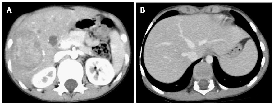
Figure 1 Contrasted-enhanced computed tomography scan of the abdomen.
A: Arterial phase: a 6.3 cm × 5.1 cm × 5.5 cm, relatively well-defined, hypo-dense lesion with internal enhancement involving the right hepatic lobe (segments# V, VI and VII); B: Delayed venous phase: the right hepatic vein was thrombosed, whereas the middle and left hepatic veins, as well as the inferior vena cava, were patent.
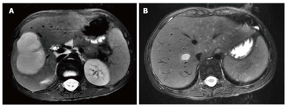
Figure 2 T2-attenuated magnetic resonance imaging scan of the abdomen.
A: A 4.7 cm × 4.7 cm × 6.6 cm, contrast-enhancing, hyper-intensive, well-defined, and moderate- to large-sized lesion involving the right hepatic lobe (segments# V, VI and VII); B: There was extension of the known hepatic IPT lesion into the path of the right hepatic vein.
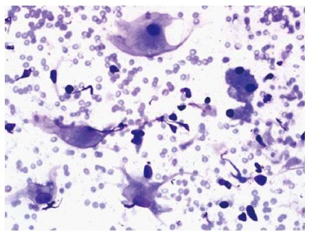
Figure 3 Fine-needle aspiration of the focal hepatic lesion.
The smear contains a mixture of benign hepatocytes with histiocytes (Diff Quick stain, magnification power: × 40).
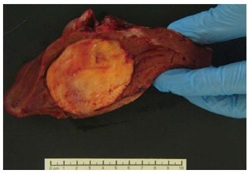
Figure 4 Gross picture of the right hepatic lobectomy.
There is a focal, soft, fleshy, yellow-white, and tanned nodule measuring 8 cm × 6 cm × 5 cm.
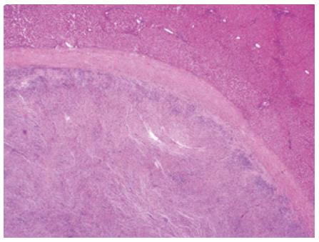
Figure 5 Focal hepatic lesion was surrounded by a well-demarcated thin capsule (HE stain, magnification power: × 20).
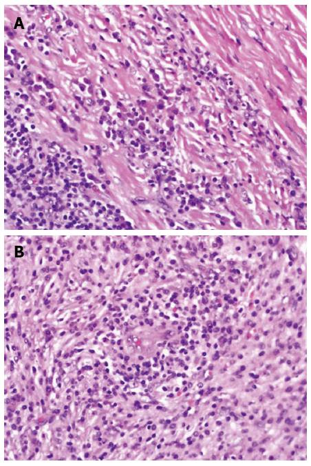
Figure 6 Microscopic picture of the focal hepatic lesion displaying a mixture of inflammatory cells (histiocytes, plasma cells, mature lymphocytes, and occasional multinucleated giant cells) in a background of dense fibrous tissues.
The inflammatory cells were mostly concentrated around the sub-capsular area at the periphery (A) and around the blood vessels (B) (HE stain, magnification power: × 40).
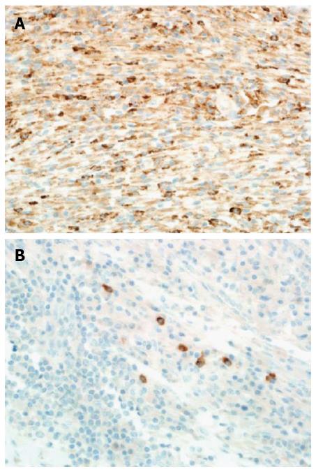
Figure 7 Immunohistochemical analysis of the focal hepatic lesion.
A: All histiocytes in the lesion stained diffusely positive for CD-68 (HE stain, magnification power: × 40). B: A few plasma cells in the lesion stained focally positive for IgG4 (IgG4 stain, magnification power: × 40).
- Citation: Al-Hussaini H, Azouz H, Abu-Zaid A. Hepatic inflammatory pseudotumor presenting in an 8-year-old boy: A case report and review of literature. World J Gastroenterol 2015; 21(28): 8730-8738
- URL: https://www.wjgnet.com/1007-9327/full/v21/i28/8730.htm
- DOI: https://dx.doi.org/10.3748/wjg.v21.i28.8730


