Copyright
©The Author(s) 2015.
World J Gastroenterol. Jul 28, 2015; 21(28): 8629-8635
Published online Jul 28, 2015. doi: 10.3748/wjg.v21.i28.8629
Published online Jul 28, 2015. doi: 10.3748/wjg.v21.i28.8629
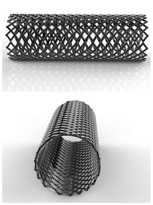
Figure 1 Example of a conventional stent.
A conventional stent designed for a rabbit’s esophagus (Shandong Provincial Institute of Medical Instruments, Jinan, China). This type of stent can remold and maintain luminal patency through a long-term dilation effect, but its radial force cannot be changed and may impede stent removal.
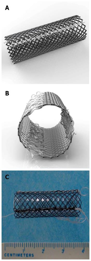
Figure 2 Newly designed detachable “pieced” stent.
A and B: Schematic drawings of the detachable “pieced” stent which contains three pieces of curved nickel-titanium mesh connected with surgical suture lines; C: Actual detachable “pieced” stent (10 mm diameter and 30 mm length) used for rabbit experiments.
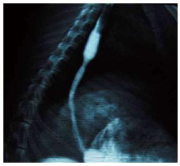
Figure 3 Esophagographic examination of the stricture modeling.
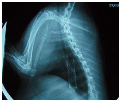
Figure 4 Stent implantation in the esophagus under fluoroscopy.
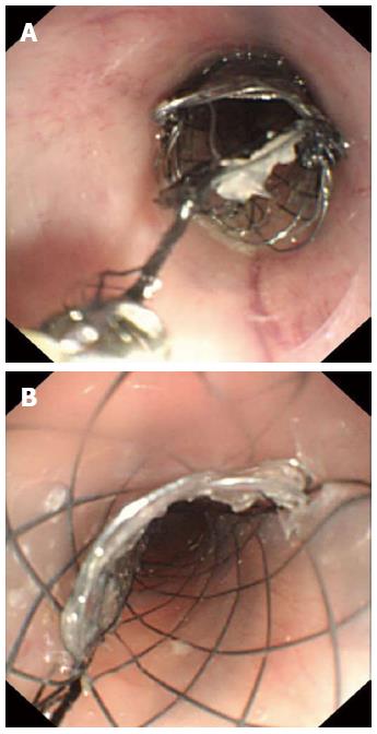
Figure 5 Illustrations of the study stent extraction.
A: The slipknots of surgical suture lines were unfastened successively after biopsy forceps pulled the string; B: Three pieces of nickel-titanium mesh connected by surgical suture lines subsequently detached.
- Citation: Liu J, Shang L, Liu JY, Qin CY. Newly designed “pieced” stent in a rabbit model of benign esophageal stricture. World J Gastroenterol 2015; 21(28): 8629-8635
- URL: https://www.wjgnet.com/1007-9327/full/v21/i28/8629.htm
- DOI: https://dx.doi.org/10.3748/wjg.v21.i28.8629


