Copyright
©The Author(s) 2015.
World J Gastroenterol. Jul 21, 2015; 21(27): 8458-8461
Published online Jul 21, 2015. doi: 10.3748/wjg.v21.i27.8458
Published online Jul 21, 2015. doi: 10.3748/wjg.v21.i27.8458
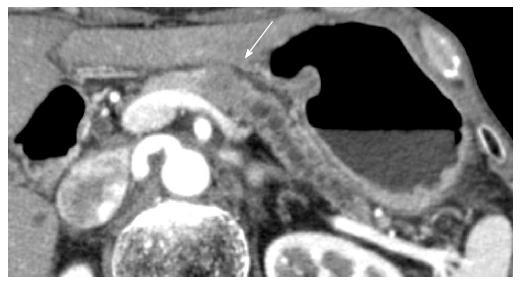
Figure 1 Computed tomography showing a 20-mm mass lesion in the pancreatic body (arrow).
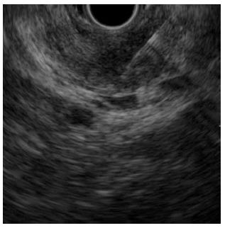
Figure 2 Endoscopic ultrasonography-guided fine needle aspiration of the 20-mm hypoechoic mass lesion was performed using a 22-G needle.
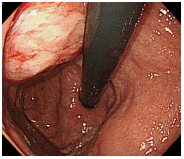
Figure 3 Submucosal tumor at the gastric body increased in size at 28 mo after surgery.
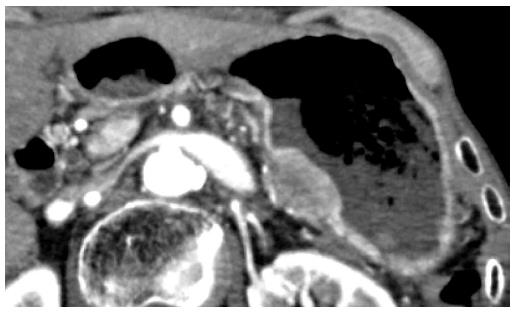
Figure 4 Computed tomography shows the submucosal tumor located adjacent to the pancreatic resection site.
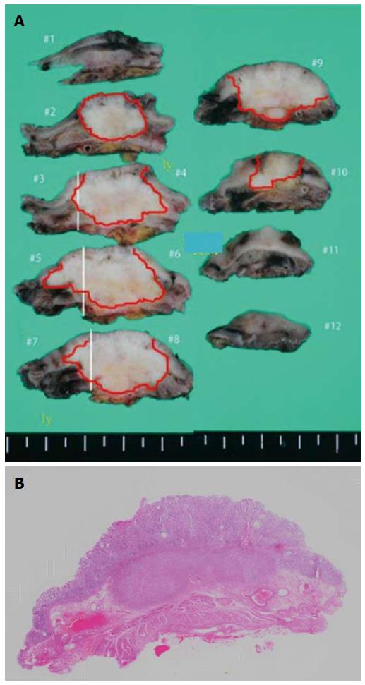
Figure 5 Resected specimen of the needle tract seeding in the stomach.
The tumor is 25 mm × 25 mm, with a whitish appearance (A). The tumor is located in the submucosal layer (B). Hematoxylin and eosin staining.
- Citation: Tomonari A, Katanuma A, Matsumori T, Yamazaki H, Sano I, Minami R, Sen-yo M, Ikarashi S, Kin T, Yane K, Takahashi K, Shinohara T, Maguchi H. Resected tumor seeding in stomach wall due to endoscopic ultrasonography-guided fine needle aspiration of pancreatic adenocarcinoma. World J Gastroenterol 2015; 21(27): 8458-8461
- URL: https://www.wjgnet.com/1007-9327/full/v21/i27/8458.htm
- DOI: https://dx.doi.org/10.3748/wjg.v21.i27.8458


