Copyright
©The Author(s) 2015.
World J Gastroenterol. Jun 14, 2015; 21(22): 7059-7064
Published online Jun 14, 2015. doi: 10.3748/wjg.v21.i22.7059
Published online Jun 14, 2015. doi: 10.3748/wjg.v21.i22.7059
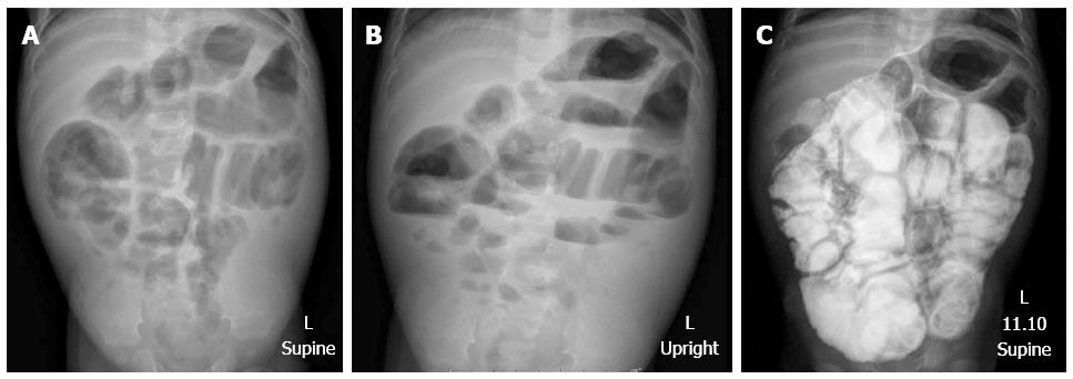
Figure 1 Plain abdominal radiographs showed diffused dilatation of small bowel loops.
A: Supine; B: Upright; C: Small bowel follow-through showed dilatation with thickening fold of almost the entire small bowel, from duodenum to ileum.
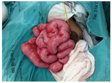
Figure 2 Exploratory laparotomy revealed thickening of the short small bowel.
The bowel was pale, thickened, and an inflamed Tenia coli-like line was noted on the antimesenteric side from the duodenojejunal junction to 15 cm above the ileocecal valve.
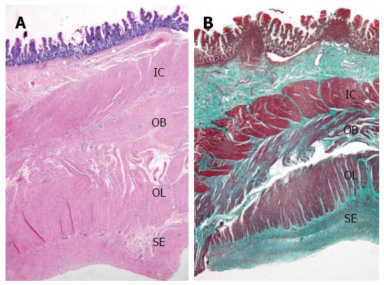
Figure 3 Full-thickness biopsy from distal ileum.
A: (HE, 20×) Hypertrophic muscularis propria with abnormal layering into 3 layers (IC: Inner circular; OB: Additional oblique; OL: Outer longitudinal); B: Delicate interstitial fibrosis and serosal muscularization (SE) are highlighted by Masson’s trichrome staining (20×).
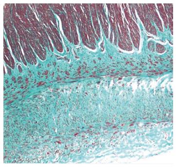
Figure 4 Serosal aberrant muscularization into three bizarre layers of smooth muscle (Masson’s trichrome, 100×).
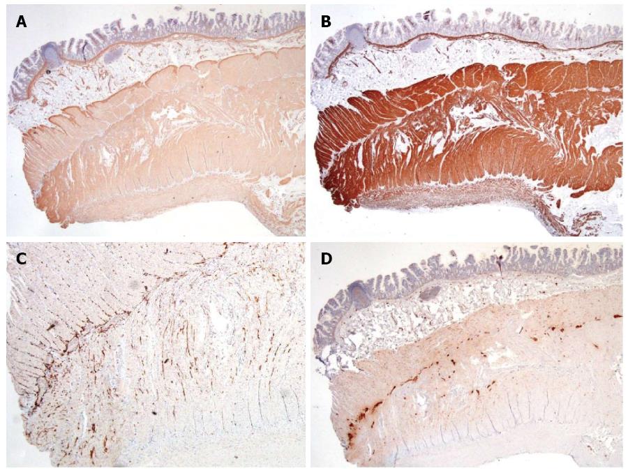
Figure 5 Immunohistochemical study.
A: Smooth muscle α-actin; B: Desmin were strongly expressed in all layers of smooth muscle; C: CD117 showed interstitial cells of Cajal network; D: S100 highlighted Auerbach’s neural plexuses; (20×).
- Citation: Angkathunyakul N, Treepongkaruna S, Molagool S, Ruangwattanapaisarn N. Abnormal layering of muscularis propria as a cause of chronic intestinal pseudo-obstruction: A case report and literature review. World J Gastroenterol 2015; 21(22): 7059-7064
- URL: https://www.wjgnet.com/1007-9327/full/v21/i22/7059.htm
- DOI: https://dx.doi.org/10.3748/wjg.v21.i22.7059


