Copyright
©The Author(s) 2015.
World J Gastroenterol. Jun 7, 2015; 21(21): 6479-6490
Published online Jun 7, 2015. doi: 10.3748/wjg.v21.i21.6479
Published online Jun 7, 2015. doi: 10.3748/wjg.v21.i21.6479
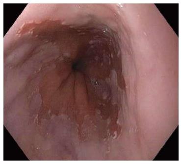
Figure 1 Endoscopic picture of gastroesophageal junction with Barrett’s esophagus.
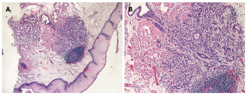
Figure 2 Photomicrograph of endoscopic mucosal resection specimen of neosquamous mucosa showing buried (subsquamous) adenocarcinoma.
A: Low magnification of entire endoscopic mucosal resection tissue; B: Medium magnification of submucosal lesion.
- Citation: Halland M, Katzka D, Iyer PG. Recent developments in pathogenesis, diagnosis and therapy of Barrett's esophagus. World J Gastroenterol 2015; 21(21): 6479-6490
- URL: https://www.wjgnet.com/1007-9327/full/v21/i21/6479.htm
- DOI: https://dx.doi.org/10.3748/wjg.v21.i21.6479


