Copyright
©The Author(s) 2015.
World J Gastroenterol. May 28, 2015; 21(20): 6391-6397
Published online May 28, 2015. doi: 10.3748/wjg.v21.i20.6391
Published online May 28, 2015. doi: 10.3748/wjg.v21.i20.6391
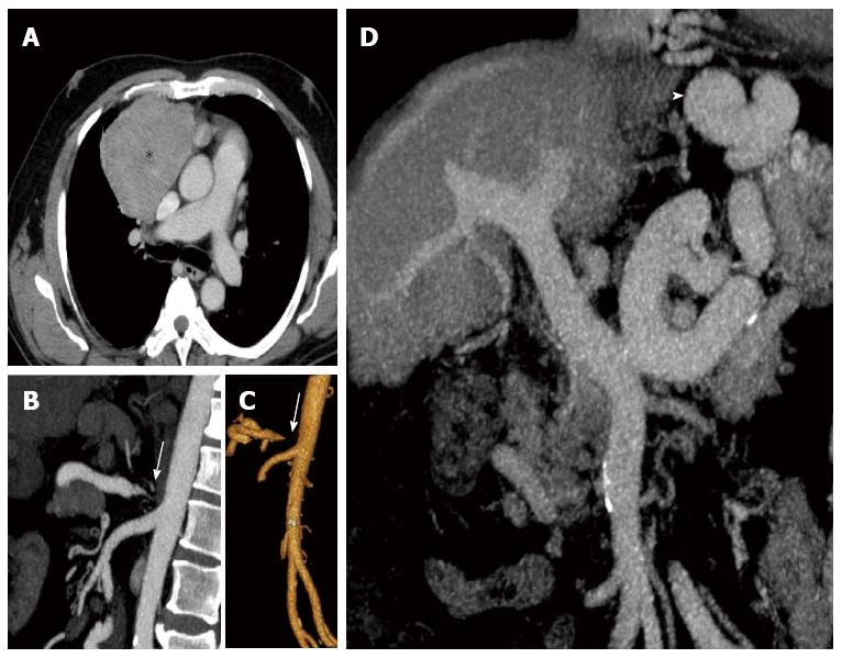
Figure 1 Axial, coronal and sagittal reconstructions of enhanced-computed tomography scan of the patient.
A: Shows the anterior mediastinal mass (Asterisk) compressing and displacing the adjacent structures (after biopsy, the patient was diagnosed with thymoma). To reverse the thrombocytopenia to initiate chemotherapy treatment, the first option was splenic artery embolization, but this was contraindicated due to severe stenosis of the celiac trunk [arrows in (B) and (C)]; D: Cirrhotic liver, and the large portal trunk caliber, all characteristics of severe portal hypertension secondary to schistosomiasis. Also of note, the large sized collateral circulation (arrowhead in D) common to this profile.
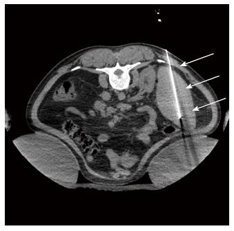
Figure 2 Axial non-enhanced computed tomography scan guiding the splenic radiofrequency ablation, with the patient in the prone position.
Note the “single” probe (arrow) in the middle/ lower third of the spleen. where the ablation zones were concentrated.
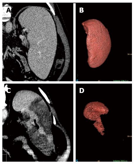
Figure 3 Coronal reformatted contrast-enhanced computed tomography and 3D volumetric reconstruction of the spleen before (A and B) and after (C and D) radiofrequency ablation.
Observe the homogeneous splenic parenchyma (A) showing a volume of 1296.2 cc (B) before treatment. After ablation, note the heterogeneity of the spleen (C), with multiple areas of coagulative necrosis mainly concentrated in the middle and lower thirds, saving the splenic hilum and its upper third. After ablation, the volumetric 3D reconstruction confirmed treatment success, leaving approximately 30% of viable parenchyma (volume of 359.5 cc) (D).
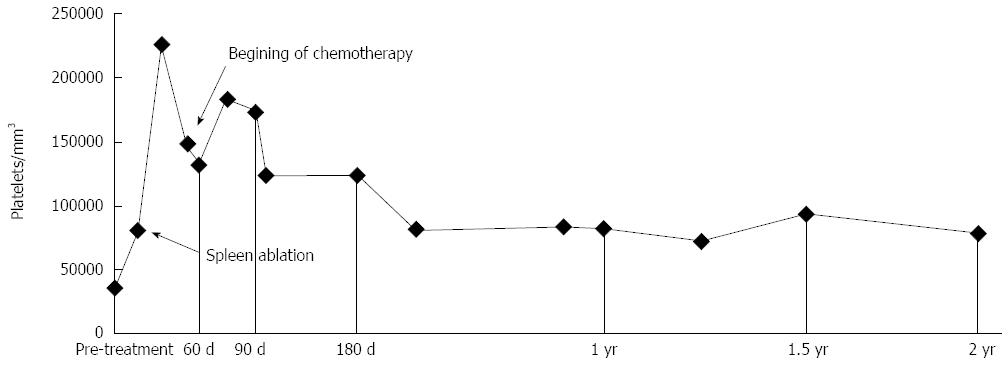
Figure 4 Graph above shows good recovery of platelet levels in a short period of time following the procedure, maintaining adequate levels during follow-up, and allowing chemotherapy to be conducted.
- Citation: Martins GLP, Bernardes JPG, Rovella MS, Andrade RG, Viana PCC, Herman P, Cerri GG, Menezes MR. Radiofrequency ablation for treatment of hypersplenism: A feasible therapeutic option. World J Gastroenterol 2015; 21(20): 6391-6397
- URL: https://www.wjgnet.com/1007-9327/full/v21/i20/6391.htm
- DOI: https://dx.doi.org/10.3748/wjg.v21.i20.6391


