Copyright
©The Author(s) 2015.
World J Gastroenterol. May 28, 2015; 21(20): 6391-6397
Published online May 28, 2015. doi: 10.3748/wjg.v21.i20.6391
Published online May 28, 2015. doi: 10.3748/wjg.v21.i20.6391
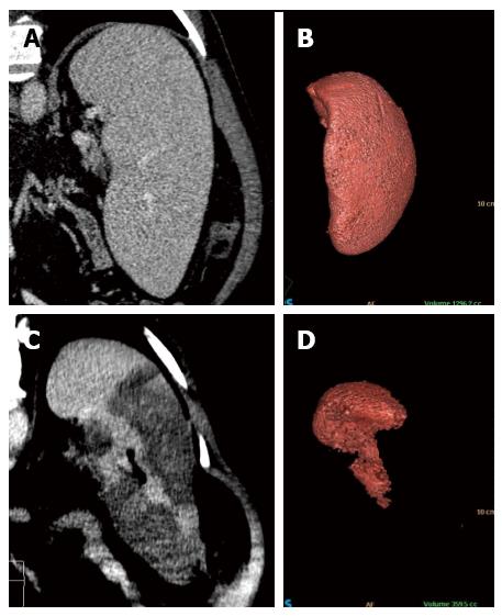
Figure 3 Coronal reformatted contrast-enhanced computed tomography and 3D volumetric reconstruction of the spleen before (A and B) and after (C and D) radiofrequency ablation.
Observe the homogeneous splenic parenchyma (A) showing a volume of 1296.2 cc (B) before treatment. After ablation, note the heterogeneity of the spleen (C), with multiple areas of coagulative necrosis mainly concentrated in the middle and lower thirds, saving the splenic hilum and its upper third. After ablation, the volumetric 3D reconstruction confirmed treatment success, leaving approximately 30% of viable parenchyma (volume of 359.5 cc) (D).
- Citation: Martins GLP, Bernardes JPG, Rovella MS, Andrade RG, Viana PCC, Herman P, Cerri GG, Menezes MR. Radiofrequency ablation for treatment of hypersplenism: A feasible therapeutic option. World J Gastroenterol 2015; 21(20): 6391-6397
- URL: https://www.wjgnet.com/1007-9327/full/v21/i20/6391.htm
- DOI: https://dx.doi.org/10.3748/wjg.v21.i20.6391


