Copyright
©The Author(s) 2015.
World J Gastroenterol. May 28, 2015; 21(20): 6206-6214
Published online May 28, 2015. doi: 10.3748/wjg.v21.i20.6206
Published online May 28, 2015. doi: 10.3748/wjg.v21.i20.6206
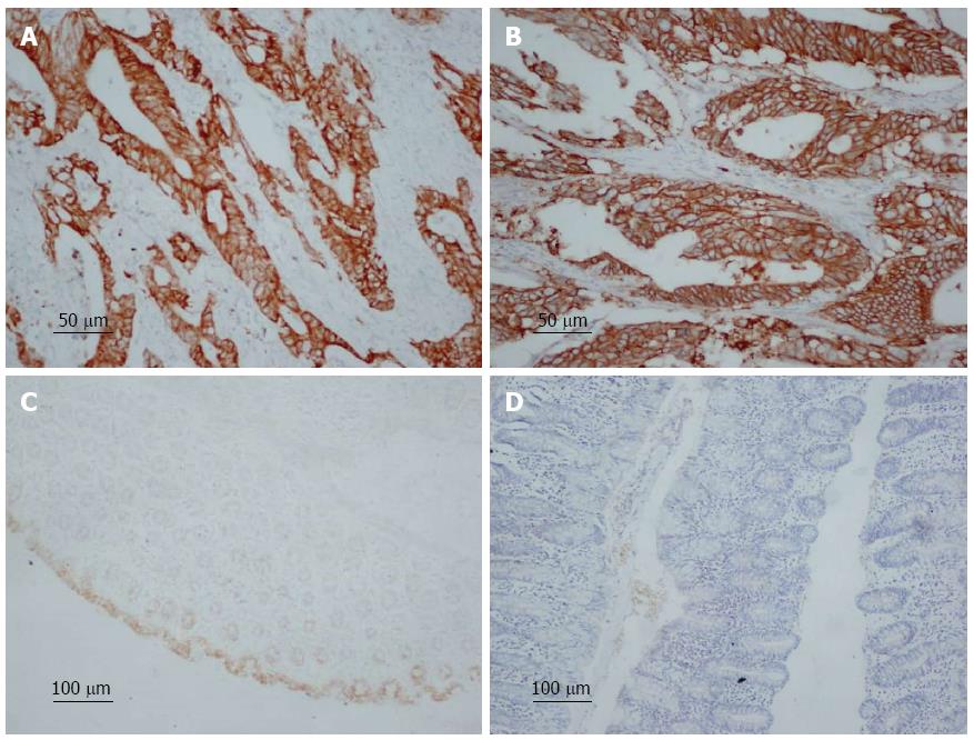
Figure 1 Immunohistochemical staining of COX-2 and HER-2 in colorectal cancer and normal colorectal tissue.
A: COX-2-positive expression in colorectal cancer, magnification × 200; B: HER-2 positive expression in colorectal cancer, magnification × 200; C: COX-2-negative expression in normal colorectal tissue, magnification × 100; D: HER-2-negative expression in normal colorectal tissue, magnification × 100.
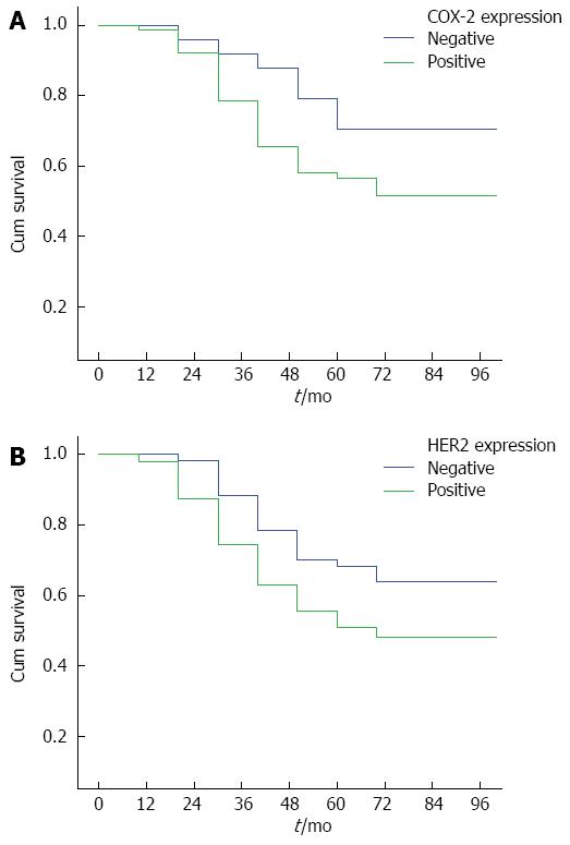
Figure 2 Survival curves for patients with colorectal cancer.
A: Patients with positive and negative COX-2 expression; B: Patients with positive and negative HER-2 expression.
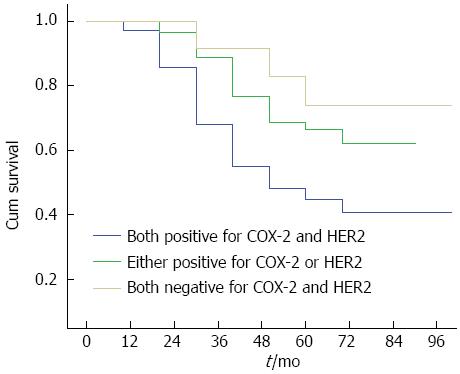
Figure 3 Survival curves of colorectal cancer patients positive for both COX-2 and HER-2, positive for either of them, and negative for both.
Compared with patients positive for both markers, patients negative for both had better survival.
- Citation: Wu QB, Sun GP. Expression of COX-2 and HER-2 in colorectal cancer and their correlation. World J Gastroenterol 2015; 21(20): 6206-6214
- URL: https://www.wjgnet.com/1007-9327/full/v21/i20/6206.htm
- DOI: https://dx.doi.org/10.3748/wjg.v21.i20.6206


