Copyright
©The Author(s) 2015.
World J Gastroenterol. May 14, 2015; 21(18): 5630-5634
Published online May 14, 2015. doi: 10.3748/wjg.v21.i18.5630
Published online May 14, 2015. doi: 10.3748/wjg.v21.i18.5630
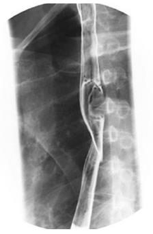
Figure 1 Representative findings from esophageal barium meal of an esophageal gastrointestinal stromal tumor.
It shows a larger tumor in the esophagus.
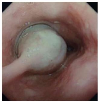
Figure 2 Representative findings from esophagoscopy.
A large and regular tumor in the esophagus.
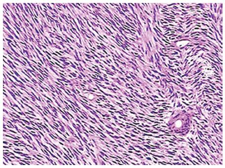
Figure 3 Representative findings from pathological examination of esophageal gastrointestinal stromal tumors.
The hematoxylin-eosin stained gastrointestinal stromal tumor shows a rich variety of spindle cells (× 100 magnification).
- Citation: Zhang FB, Shi HC, Shu YS, Shi WP, Lu SC, Zhang XY, Tu SS. Diagnosis and surgical treatment of esophageal gastrointestinal stromal tumors. World J Gastroenterol 2015; 21(18): 5630-5634
- URL: https://www.wjgnet.com/1007-9327/full/v21/i18/5630.htm
- DOI: https://dx.doi.org/10.3748/wjg.v21.i18.5630


