Copyright
©The Author(s) 2015.
World J Gastroenterol. May 14, 2015; 21(18): 5560-5567
Published online May 14, 2015. doi: 10.3748/wjg.v21.i18.5560
Published online May 14, 2015. doi: 10.3748/wjg.v21.i18.5560
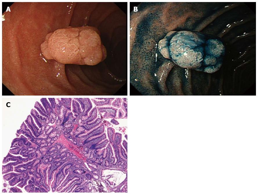
Figure 1 Case of duodenal adenoma.
A: A whitish polypoid lesion is observed in the second portion of the duodenum; B: Chromoendoscopy clarifies the presence of lobulation. The lesion was diagnosed as a 0-Isp type adenoma; C: Histology of the biopsy specimen confirms a non-carcinomatous lesion, and the final diagnosis is a low-grade adenoma.
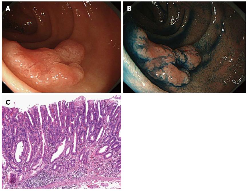
Figure 2 A case of duodenal carcinoma.
A: A slightly elevated lesion with a reddish depression is observed in the second portion of the duodenum; B: Chromoendoscopy clarifies the presence of depressions. The lesion was diagnosed as a 0-IIa+IIc type intramucosal carcinoma; C: The final diagnosis is well-differentiated intramucosal adenocarcinoma.
- Citation: Kakushima N, Kanemoto H, Sasaki K, Kawata N, Tanaka M, Takizawa K, Imai K, Hotta K, Matsubayashi H, Ono H. Endoscopic and biopsy diagnoses of superficial, nonampullary, duodenal adenocarcinomas. World J Gastroenterol 2015; 21(18): 5560-5567
- URL: https://www.wjgnet.com/1007-9327/full/v21/i18/5560.htm
- DOI: https://dx.doi.org/10.3748/wjg.v21.i18.5560


