Copyright
©The Author(s) 2015.
World J Gastroenterol. May 14, 2015; 21(18): 5548-5554
Published online May 14, 2015. doi: 10.3748/wjg.v21.i18.5548
Published online May 14, 2015. doi: 10.3748/wjg.v21.i18.5548
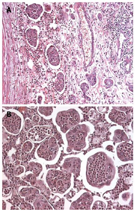
Figure 1 Invasive micropapillary carcinoma characterized by cell clusters surrounded by lacunar spaces and fibrous stroma (A) and several micropapillae infiltrated by numerous neutrophils.
A: HE staining, magnification × 100; B: HE staining, magnification × 400.
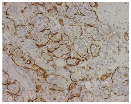
Figure 2 Micropapillary clusters were characteristically MUC-1 immunoreactive on the stroma-facing surface (“inside-out” pattern).
Magnification × 200.
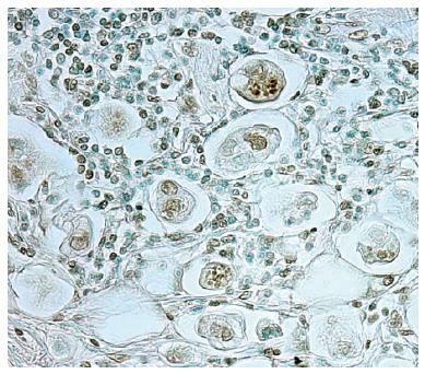
Figure 3 Intraepithelial neutrophils showing TUNEL-positivity.
Magnification × 200.
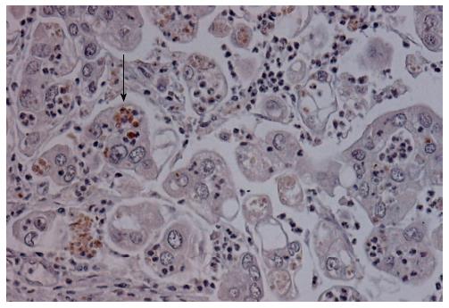
Figure 4 Neutrophils showing cytoplasmic immunoreactivity for caspase-3 are found within cytoplasmic vacuoles of tumor cells (arrow).
Magnification × 200.
- Citation: Barresi V, Branca G, Ieni A, Rigoli L, Tuccari G, Caruso RA. Phagocytosis (cannibalism) of apoptotic neutrophils by tumor cells in gastric micropapillary carcinomas. World J Gastroenterol 2015; 21(18): 5548-5554
- URL: https://www.wjgnet.com/1007-9327/full/v21/i18/5548.htm
- DOI: https://dx.doi.org/10.3748/wjg.v21.i18.5548


