Copyright
©The Author(s) 2015.
World J Gastroenterol. May 14, 2015; 21(18): 5488-5495
Published online May 14, 2015. doi: 10.3748/wjg.v21.i18.5488
Published online May 14, 2015. doi: 10.3748/wjg.v21.i18.5488
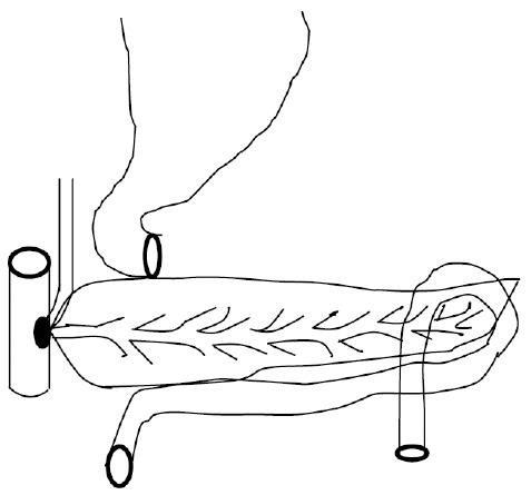
Figure 1 Technique of pancreas-sparing duodenectomy.
En bloc resection of the biliopancreatic junction and descending segment of duodenum.
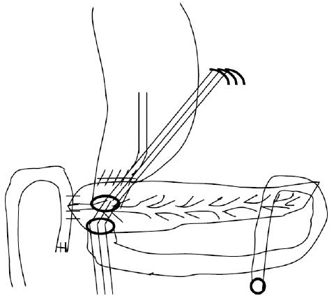
Figure 2 Pancreaticojejunostomy, choledochojejunostomy, and end-to-end anastomosis of the duodenum are performed with pyloroplasty.
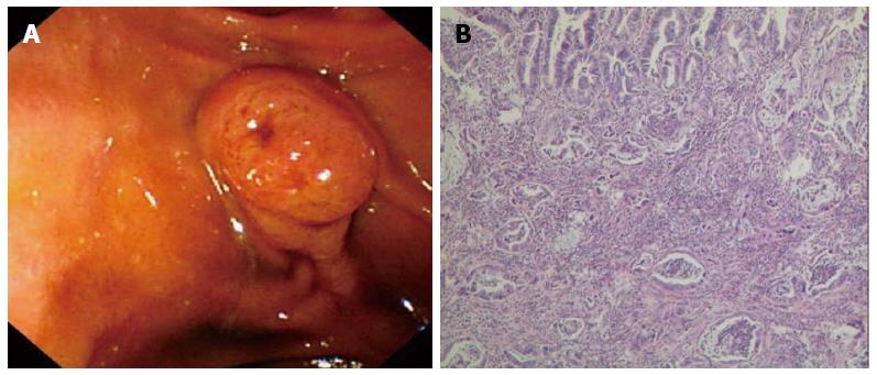
Figure 3 Duodenoscopic view of ampullary adenocarcinoma (A) and histopathological features of mixed type ampullary adenocarcinoma (B).
A: An exposed-type tumor mass at the ampulla of Vater, with a normal ampullary orifice; B: Histopathological features of mixed type ampullary adenocarcinoma. Both intestinal and pancreatobiliary growth patterns were present (HE staining, × 200).
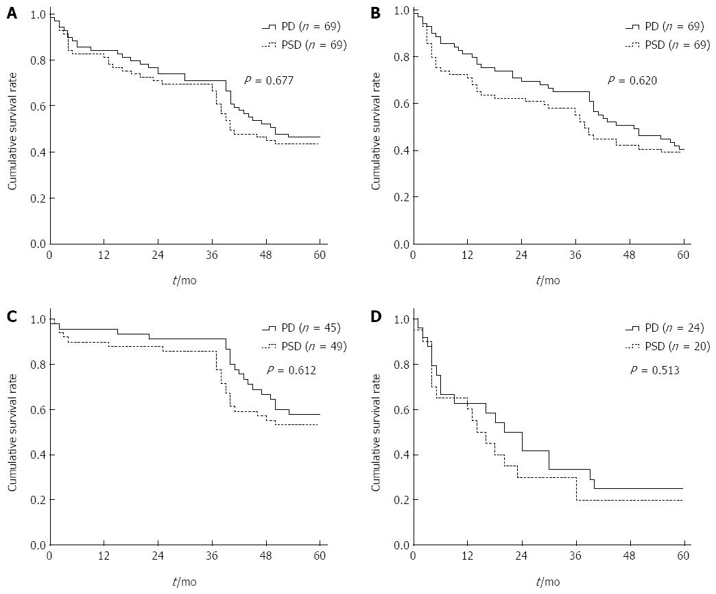
Figure 4 Kaplan-Meier survival analysis of all patients.
A: Overall survival (P = 0.677); B: Disease-free survival (P = 0.62); C: Overall survival of N0 patients (P = 0.612); D: Overall survival of N1 patients (P = 0.513).
- Citation: Liu B, Li J, Zhang YJ, Yan LN, You SY, Lau WY, Sun HR, Yan SY, Wang ZQ. Pancreas-sparing duodenectomy with regional lymph node dissection for early-stage ampullary carcinoma: A case control study using propensity scoring methods. World J Gastroenterol 2015; 21(18): 5488-5495
- URL: https://www.wjgnet.com/1007-9327/full/v21/i18/5488.htm
- DOI: https://dx.doi.org/10.3748/wjg.v21.i18.5488


