Copyright
©The Author(s) 2015.
World J Gastroenterol. Apr 28, 2015; 21(16): 4829-4839
Published online Apr 28, 2015. doi: 10.3748/wjg.v21.i16.4829
Published online Apr 28, 2015. doi: 10.3748/wjg.v21.i16.4829
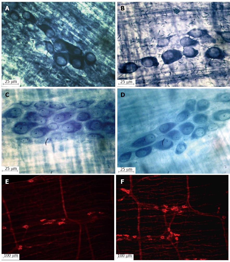
Figure 1 Photomicrograph of the myenteric ganglia in the jejunum of healthy (A, C and E) and infected rats (B, D and F); NADH-diaphorase (A and B); Giemsa (C and D).
NOS-IR (E and F) showing the increase in the nitrergic myenteric neuron population in rats infected with oocysts of the ME-49 genotype II (F) strain of Toxoplasma gondii.
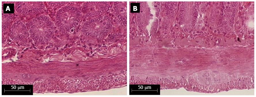
Figure 2 External muscle layer thickness of the jejunal wall of healthy (A) and infected rats (B) with the ME-49 genotype II strain of Toxoplasma gondii, colored by the HE histological technique.
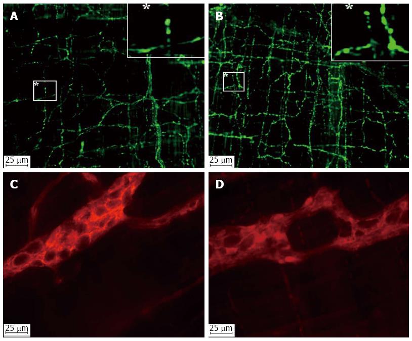
Figure 3 Photomicrographs of the VIPergic fibers (A and B) and enteric glial cells (C and D) in the jejunal myenteric plexus of healthy (A and C) and infected (B and D) rats with the ME-49 genotype II strain of Toxoplasma gondii.
-
Citation: Araújo EJA, Zaniolo LM, Vicentino SL, Góis MB, Zanoni JN, Silva AVD, Sant’Ana DMG.
Toxoplasma gondii causes death and plastic alteration in the jejunal myenteric plexus. World J Gastroenterol 2015; 21(16): 4829-4839 - URL: https://www.wjgnet.com/1007-9327/full/v21/i16/4829.htm
- DOI: https://dx.doi.org/10.3748/wjg.v21.i16.4829


