Copyright
©The Author(s) 2015.
World J Gastroenterol. Apr 14, 2015; 21(14): 4408-4412
Published online Apr 14, 2015. doi: 10.3748/wjg.v21.i14.4408
Published online Apr 14, 2015. doi: 10.3748/wjg.v21.i14.4408
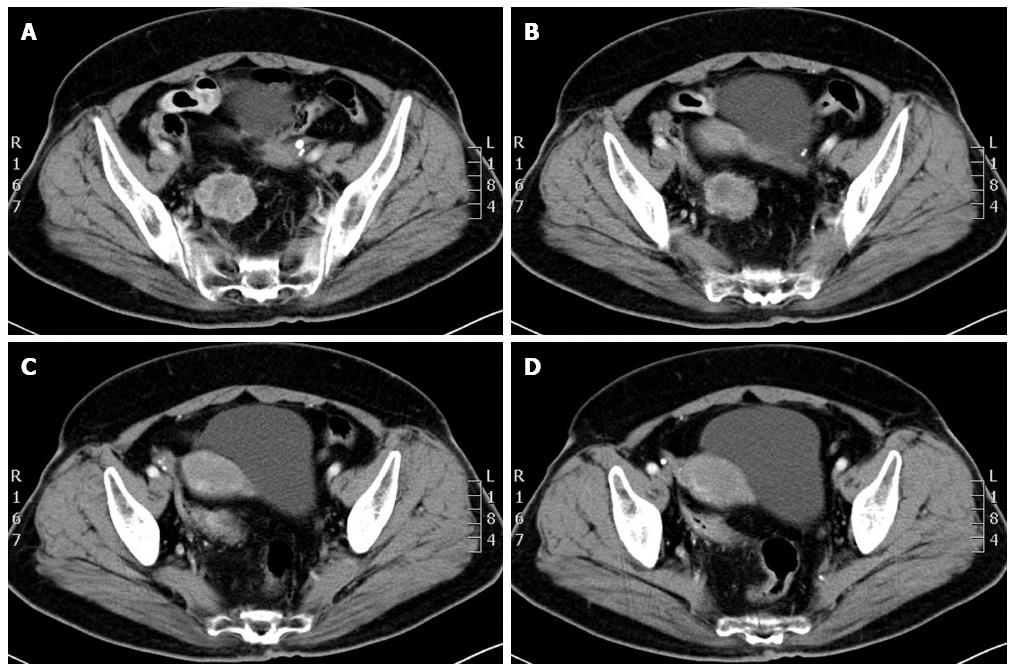
Figure 1 Contrast enhanced computed tomography showing a pelvic soft-tissue lesion which had heterogeneous density in the venous and delay phases, respectively.
Figure 2 Mesorectum and portion of the rectum.
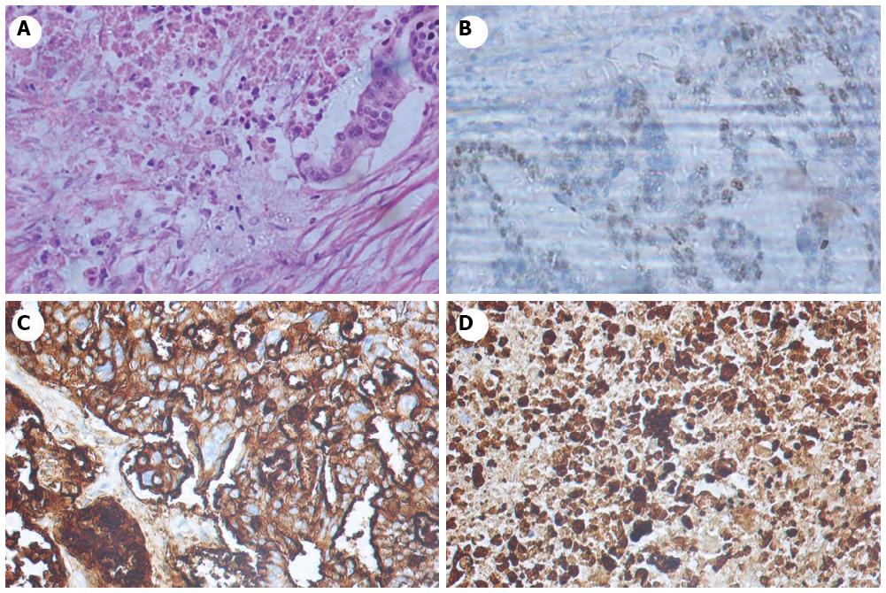
A: Microscopically, the tumor consisted of single-file stands of infiltrating tumor cells with the presence of signet-ring cells (hematoxylin-eosin staining, magnification × 400). Immunohistochemical staining of tumor cells; B: Positive for estrogen receptor protein; C: Strongly positive for CA-125; D: Strongly positive for CK7.
- Citation: Xue F, Liu ZL, Zhang Q, Kong XN, Liu WZ. Mesorectum localization as a special kind of rectal metastasis from breast cancer. World J Gastroenterol 2015; 21(14): 4408-4412
- URL: https://www.wjgnet.com/1007-9327/full/v21/i14/4408.htm
- DOI: https://dx.doi.org/10.3748/wjg.v21.i14.4408


