Copyright
©The Author(s) 2015.
World J Gastroenterol. Apr 14, 2015; 21(14): 4402-4407
Published online Apr 14, 2015. doi: 10.3748/wjg.v21.i14.4402
Published online Apr 14, 2015. doi: 10.3748/wjg.v21.i14.4402
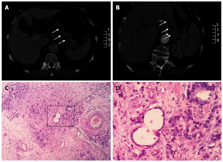
Figure 1 Upper abdomen computed tomography scan.
A: Plain computed tomography (CT) indicated a localized, irregularly-shaped mass in the stomach; however, no obvious lymph node metastasis was observed; B: Contrast-enhanced CT scan revealed inhomogeneous enhancement of the gastric wall lesion; C, D. Biopsy results pathologically confirmed the diagnosis of a poorly differentiated adenocarcinoma. Microscopic examination revealed obvious atypical nuclei and large, bizarre, multinucleated cell mitosis. Generally, the normal structure of gastric gland was lost. (Hematoxylin and eosin staining, C: Magnification × 100; D: Magnification × 400).
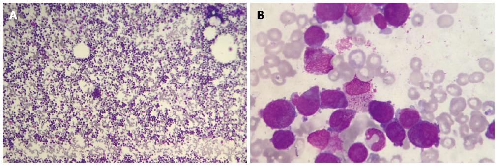
Figure 2 Peripheral blood smear, bone marrow biopsy and immunohistochemistry results revealed that the total number of primitive cells plus immature leukemia cells account up to 91.
5% (2% + 89.5%) of the total cell population. Leukemic hiatus was detected, which supports the diagnosis of acute myeloid leukemia (M3a, APL). (Hematoxylin and eosin staining, A: Magnification × 10; B: Magnification × 400).
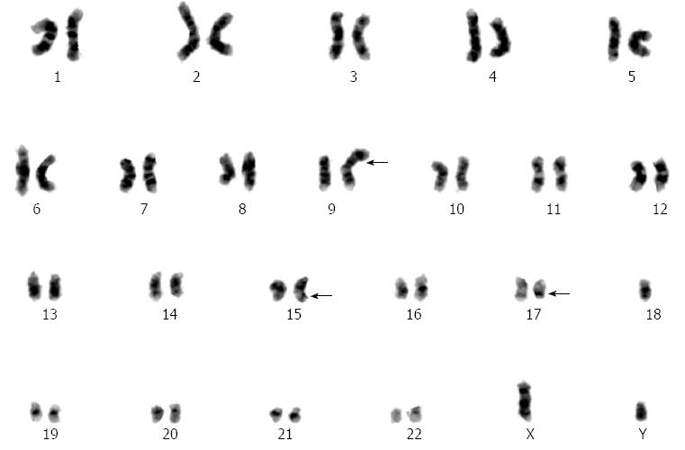
Figure 3 Chromosomal analysis via karyotype revealed 46 chromosomes with abnormities present in chromosomes 9, 15, and 17.
Unbalanced translocation occurred in chromosomes 15 and 17. 46,XY,t (15;17)(q22;q21)[15]/46,idem,-9,+add(9)(p?22)[2]/46,XY[3].
- Citation: Zhang YC, Zhou YQ, Yan B, Shi J, Xiu LJ, Sun YW, Liu X, Qin ZF, Wei PK, Li YJ. Secondary acute promyelocytic leukemia following chemotherapy for gastric cancer: A case report. World J Gastroenterol 2015; 21(14): 4402-4407
- URL: https://www.wjgnet.com/1007-9327/full/v21/i14/4402.htm
- DOI: https://dx.doi.org/10.3748/wjg.v21.i14.4402


