Copyright
©The Author(s) 2015.
World J Gastroenterol. Apr 14, 2015; 21(14): 4397-4401
Published online Apr 14, 2015. doi: 10.3748/wjg.v21.i14.4397
Published online Apr 14, 2015. doi: 10.3748/wjg.v21.i14.4397
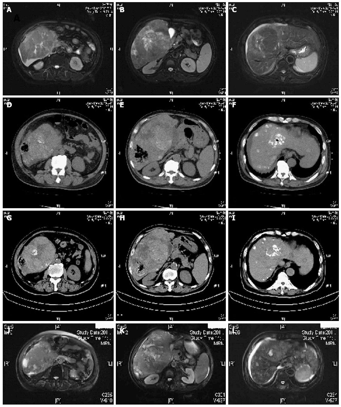
Figure 1 Magnetic resonance imaging and computed tomography findings.
A-C: Magnetic resonance imaging (MRI) images before transcatheter arterial chemoembolization (TACE); D-F: Computed tomography (CT) images showing a gas-forming liver abscess when the first TACE was done; G-I: Five week after the first TACE procedure, CT image showed that the gas-forming liver abscess became smaller; J-L: Eleven week after the first TACE procedure, MRI showed no obvious liver abscess.
-
Citation: Li JH, Yao RR, Shen HJ, Zhang L, Xie XY, Chen RX, Wang YH, Ren ZG.
Clostridium perfringens infection after transarterial chemoembolization for large hepatocellular carcinoma. World J Gastroenterol 2015; 21(14): 4397-4401 - URL: https://www.wjgnet.com/1007-9327/full/v21/i14/4397.htm
- DOI: https://dx.doi.org/10.3748/wjg.v21.i14.4397


