Copyright
©The Author(s) 2015.
World J Gastroenterol. Apr 7, 2015; 21(13): 4048-4062
Published online Apr 7, 2015. doi: 10.3748/wjg.v21.i13.4048
Published online Apr 7, 2015. doi: 10.3748/wjg.v21.i13.4048
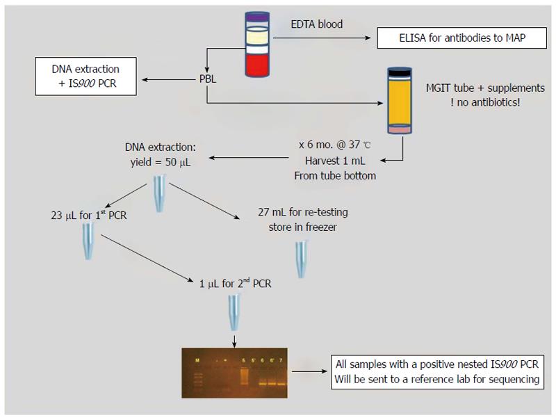
Figure 1 Schematic of sample processing and testing methods.
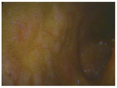
Figure 2 Terminal ileum with multiple ulcers.
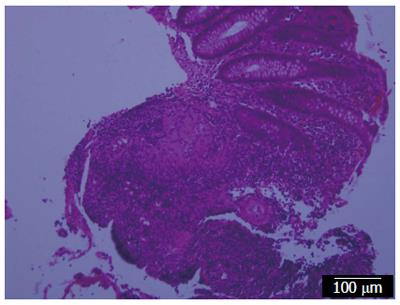
Figure 3 Biopsy from the colon showing a granuloma of Crohn’s disease.
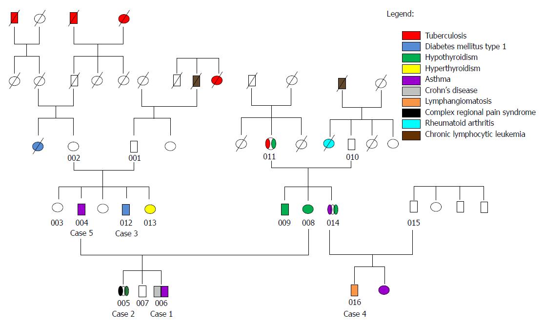
Figure 4 Family pedigree summarizing history of mycobacterial infection and other diseases of cases 1 through 5 and additional family members.
- Citation: Kuenstner JT, Chamberlin W, Naser SA, Collins MT, Dow CT, Aitken JM, Weg S, Telega G, John K, Haas D, Eckstein TM, Kali M, Welch C, Petrie T. Resolution of Crohn's disease and complex regional pain syndrome following treatment of paratuberculosis. World J Gastroenterol 2015; 21(13): 4048-4062
- URL: https://www.wjgnet.com/1007-9327/full/v21/i13/4048.htm
- DOI: https://dx.doi.org/10.3748/wjg.v21.i13.4048


